. 2016 Oct;5(5):870-882.
doi: 10.1002/mbo3.376. Epub 2016 May 10.

Zinc coordination is essential for the function and activity of the type II secretion ATPase EpsE

Affiliations

Zinc coordination is essential for the function and activity of the type II secretion ATPase EpsE

Chelsea S Rule et al. Microbiologyopen. 2016 Oct.

Abstract

The type II secretion system Eps in Vibrio cholerae promotes the extracellular transport of cholera toxin and several hydrolytic enzymes and is a major virulence system in many Gram-negative pathogens which is structurally related to the type IV pilus system. The cytoplasmic ATPase EpsE provides the energy for exoprotein secretion through ATP hydrolysis. EpsE contains a unique metal-binding domain that coordinates zinc through a tetracysteine motif (CXXCX29 CXXC), which is also present in type IV pilus assembly but not retraction ATPases. Deletion of the entire domain or substitution of any of the cysteine residues that coordinate zinc completely abrogates secretion in an EpsE-deficient strain and has a dominant negative effect on secretion in the presence of wild-type EpsE. Consistent with the in vivo data, chemical depletion of zinc from purified EpsE hexamers results in loss of in vitro ATPase activity. In contrast, exchanging the residues between the two dicysteines with those from the homologous ATPase XcpR from Pseudomonas aeruginosa does not have a significant impact on EpsE. These results indicate that, although the individual residues in the metal-binding domain are generally interchangeable, zinc coordination is essential for the activity and function of EpsE.

Keywords: ATPase; tetracysteine; type II secretion; zinc..


Figures

Figure 1
Figure 1
Structural comparison of type II/IV secretion ATPases. The structures of the Vibrio cholerae T2S ATPase EpsE (left, PDB 1P9W), the Pseudomonas aeruginosa type IV pilus retraction ATPase PilT (center, PDB 3JVV), and the Helicobacter pylori type IV secretion ATPase HP0525 (right, PDB 1G6O) are shown. N‐terminal domains are colored green, C‐terminal domains in cyan, and the CM domain in EpsE is displayed in red with zinc represented as a blue sphere. Nucleotide is shown in orange.
Figure 2
Figure 2
The EpsE CM domain is required for secretion. (A) WT and epsE::kan strains of Vibrio cholerae TRH7000 containing empty vector (−) or pMMB plasmids encoding WT or mutant EpsE variants described on the x‐axis were grown with 200 μg mL−1 carbenicillin and 10 μmol L−1 IPTG. Culture supernatants were analyzed for VesB protease activity using a cleavable fluorogenic probe as described in Experimental Procedures. All EpsE variants showed statistical significance compared to WT EpsE (P < 0.0001). (B) The same overnight culture supernatants as in A were analyzed for lipase activity as described in Experimental Procedures. All EpsE variants showed statistical significance compared to WT EpsE (P < 0.03). (C) Expression of EpsE in WT V. cholerae TRH7000, followed by epsE::kan V. cholerae containing empty vector and epsE::kan V. cholerae expressing either WT EpsE or variants of EpsE. Samples were analyzed by SDS‐PAGE and immunoblotting for EpsE. The size of EpsE and EpsE ΔCM are indicated by black arrows, and EpsE dimers by a gray arrow. WT, wild type strain.
Figure 3
Figure 3
(A) Wild‐type (WT) Vibrio cholerae TRH7000 containing pMMB plasmids encoding WT or mutant EpsE variants described on the x‐axis were grown with 200 μg mL−1 carbenicillin and 100 μmol L−1 IPTG. Samples were prepared and assayed for protease activity as in Figure 2. Assays were performed in triplicate and standard error is shown. All EpsE variants showed statistical significance compared to WT EpsE (P < 0.0001). (B) WT V. cholerae 3083 and isogenic ΔepsC containing pMMB plasmids (empty vector [−], WT EpsE, or EpsE C4xS) were grown with 200 μg mL−1 carbenicillin and 100 μmol L−1 IPTG. Cells (C) and supernatants (S) were analyzed by SDS‐PAGE and immunoblotting for cholera toxin B subunit.
Figure 4
Figure 4
Alignment of CM domains in T2S ATPase homologs. Clustal Omega alignment of CXXCX27–30CXXC motifs of T2S ATPase homologs. Asterisks below indicate residue conservation identity, and colons and periods indicate high and low levels of residue homology, respectively.
Figure 5
Figure 5
The EpsE‐XcpR CM chimera partially complements the T2S defect in epsE::kan mutants of Vibrio cholerae. (A) Vibrio cholerae TRH7000 WT, followed by epsE::kan strains containing empty vector (−) or pMMB encoding EpsE or EpsE‐XcpR CM were grown with 200 μg mL−1 carbenicillin and 10 μmol L−1 IPTG. The last bar represents WT V. cholerae expressing EpsE‐XcpR CM induced with 100 μmol L−1 IPTG to test for negative dominance. Culture supernatants were analyzed for VesB activity using a cleavable fluorogenic probe as described in Experimental Procedures. Assays were performed in triplicate and SEM is shown. No statistically significant difference between WT EpsE and EpsE‐XcpR CM was detected using a t test (P = 0.37). (B) Overnight culture supernatants were assayed for lipase activity as in Figure 2B. No statistically significant difference between WT EpsE and EpsE‐XcpR CM was detected using a t test (P = 0.064). (C) Expression of EpsE in WT V. cholerae TRH7000 (lane 1), followed by empty vector (lane 2), WT EpsE (lane 3), or EpsE‐XcpR CM expressed in epsE::kan V. cholerae (lane 4) and induced with 10 μmol L−1 IPTG. Samples were analyzed by SDS‐PAGE and immunoblotting for EpsE.
Figure 6
Figure 6
EpsE‐Hcp1 and EpsE‐XcpR CM–Hcp1 fusions support secretion in Vibrio cholerae. (A) WT and epsE::kan strains of V. cholerae TRH7000 containing empty vector (−), pMMB encoding EpsE, EpsE‐Hcp1, or EpsE‐XcpR CM–Hcp1 were grown with 200 μg mL−1 carbenicillin and 10 μmol L−1 IPTG. Supernatants were analyzed for VesB activity using a cleavable fluorogenic probe as described in Experimental Procedures. Assays were performed in triplicate and standard error is shown. (B) Lipase assays were performed on overnight culture supernatants as in Figure 2B. Assays were performed in triplicate with standard errors displayed.
Figure 7
Figure 7
Detection of full‐length EpsE‐Hcp1 fusions in Vibrio cholerae. Overnight cultures of V. cholerae TRH7000 WT or epsE::kan strains containing empty vector (−) or pMMB plasmids encoding EpsE, EpsE‐Hcp1, or EpsE‐XcpR CM–Hcp1 were grown with 200 μg mL−1 carbenicillin and 10 μmol L−1 IPTG. Cell lysates were analyzed by SDS‐PAGE and immunoblotting using α‐EpsE antibodies (left) or α‐Hcp1 antibodies (right). Molecular mass markers are indicated and the positions of EpsE and EpsE‐Hcp1 fusions are shown with arrows. Purified EpsE‐Hcp1 protein was included as a positive control.
Figure 8
Figure 8
The EpsE‐XcpR CM–Hcp1 chimera fusion maintains in vitro ATPase activity. Purified EpsE‐Hcp1 and EpsE‐XcpR CM‐Hcp1 fusions were assayed for in vitro ATPase activity as described in Experimental Procedures.
Figure 9
Figure 9
Removal of zinc results in a loss of in vitro ATPase activity and changes the migration pattern of ΔN1‐EpsE‐Hcp1. (A) Zinc release titration curve. Increasing amounts of p‐chloromercuribenzoic acid (PCMB) result in increased zinc release. (B) Treatment of ΔN1‐EpsE‐Hcp1 protein abolishes in vitro ATPase activity. Proteins were either untreated or treated with a fourfold molar excess of PCMB or mock treated for 10 min at room temperature and assayed for ATPase activity as described in Experimental Procedures. (C) Purified ΔN1‐EpsE‐Hcp1 was untreated or incubated with a fourfold molar excess of PCMB or mock treated for 10 min at room temperature. Samples were then analyzed using native‐PAGE and stained with Coomassie.
Figure 10
Figure 10
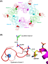
Close‐up view of residues at the base of the CM domain and potential interactions with adjacent subunits or nucleotide. (A) View along the twofold axis of the Vibrio cholerae EpsE‐8aa‐Hcp1 hexamer with C2 symmetry (Lu et al. 2013) (PDB code 4KSR). The three independent chains are related by a twofold axis and are displayed as green (chains A, A’), cyan (B, B’), and magenta (C, C’) with CM domains colored red. Arg 394 residues from each subunit are displayed as red spheres, and Leu 349 residues are displayed as spheres according to the color of the corresponding subunit. (B) Arg 394 from chain C is shown in proximity to Leu 349 of chain A’. The proximity of Arg 441 to AMPPNP is also shown, with an alpha‐helix from the EpsE C‐terminal domain in purple. Distances between possible contacts are indicated by dotted lines and labeled. Zinc is superimposed from the structure of monomeric EpsE (Robien et al. 2003) (PDB code 1P9W) and is displayed as a blue sphere.

References

    1. Abendroth, J. , Murphy P., Sandkvist M., Bagdasarian M., and Hol W. G. J.. 2005. The x‐ray structure of the type II secretion system complex formed by the N‐terminal domain of EpsE and the cytoplasmic domain of EpsL of Vibrio cholerae . J. Mol. Biol. 348:845–855. - PubMed
    1. Bischof, L. F. , Friedrich C., Harms A., Logaard‐Andersen L., and van der Does C. 2016. The type IV pilus assembly ATPase PilB of Myxococcus xanthus interacts with the inner membrane platform protein PilC and the nucleotide binding protein PilM. J. Biol. Chem. 291:6946–6957. - PMC - PubMed
    1. Camberg, J. L. , and Sandkvist M.. 2005. Molecular analysis of the Vibrio cholerae type II secretion ATPase EpsE. J. Bacteriol. 187:249–256. - PMC - PubMed
    1. Camberg, J. L. , Johnson T. L., Patrick M., Abendroth J., Hol W. G. J., and Sandkvist M.. 2007. Synergistic stimulation of EpsE ATP hydrolysis by EpsL and acidic phospholipids. EMBO J. 26:19–27. - PMC - PubMed
    1. Chiang, P. , Sampaleanu L. M., Ayers M., Pahuta M., Howell P. L., and Burrows L. L.. 2008. Functional role of conserved residues in the characteristic secretion NTPase motifs of the Pseudomonas aeruginosa type IV pilus motor proteins PilB, PilT, and PilU. Microbiology 154:114–126. - PubMed

MeSH terms

LinkOut - more resources