. 2016 May 11;12(5):e1005615.
doi: 10.1371/journal.ppat.1005615. eCollection 2016 May.

Virus Infections Incite Pain Hypersensitivity by Inducing Indoleamine 2,3 Dioxygenase

Affiliations

Virus Infections Incite Pain Hypersensitivity by Inducing Indoleamine 2,3 Dioxygenase

Lei Huang et al. PLoS Pathog. .

Abstract

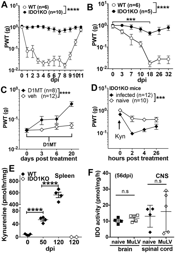
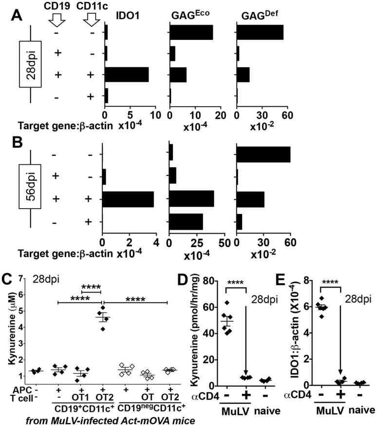
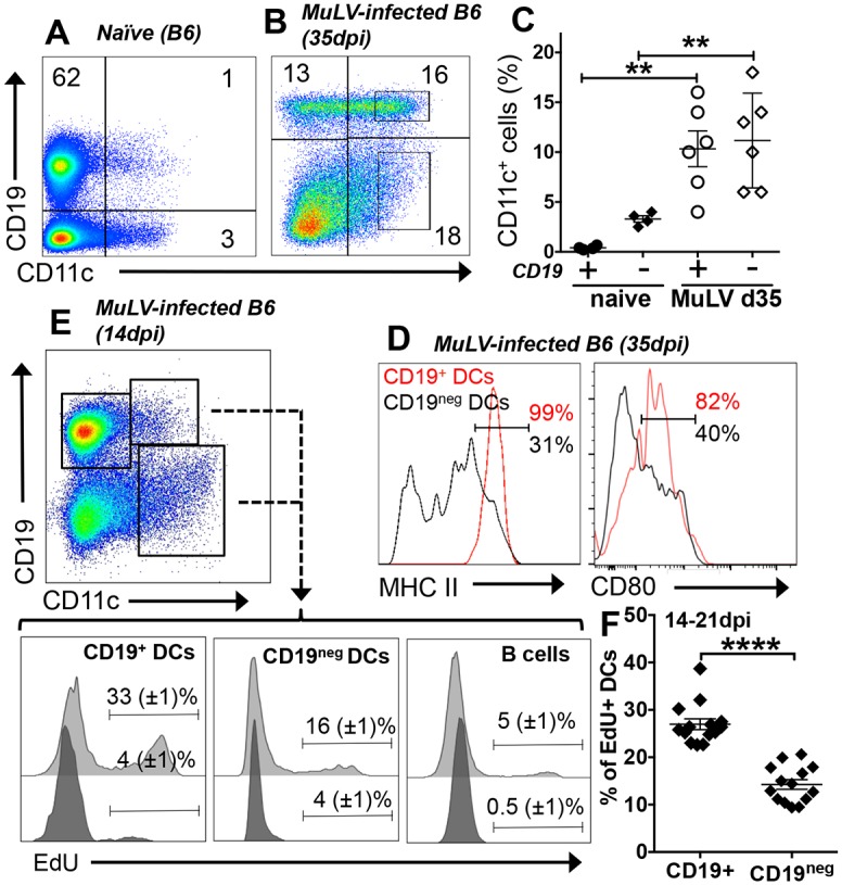
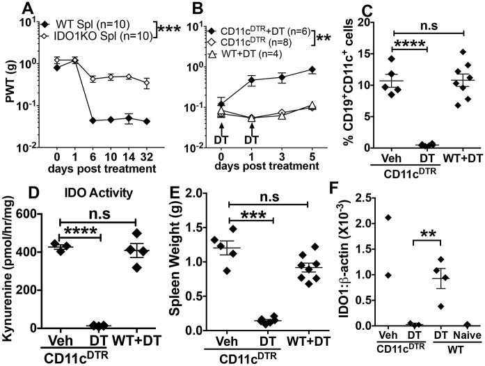
Increased pain sensitivity is a comorbidity associated with many clinical diseases, though the underlying causes are poorly understood. Recently, chronic pain hypersensitivity in rodents treated to induce chronic inflammation in peripheral tissues was linked to enhanced tryptophan catabolism in brain mediated by indoleamine 2,3 dioxygenase (IDO). Here we show that acute influenza A virus (IAV) and chronic murine leukemia retrovirus (MuLV) infections, which stimulate robust IDO expression in lungs and lymphoid tissues, induced acute or chronic pain hypersensitivity, respectively. In contrast, virus-induced pain hypersensitivity did not manifest in mice lacking intact IDO1 genes. Spleen IDO activity increased markedly as MuLV infections progressed, while IDO1 expression was not elevated significantly in brain or spinal cord (CNS) tissues. Moreover, kynurenine (Kyn), a tryptophan catabolite made by cells expressing IDO, incited pain hypersensitivity in uninfected IDO1-deficient mice and Kyn potentiated pain hypersensitivity due to MuLV infection. MuLV infection stimulated selective IDO expression by a discreet population of spleen cells expressing both B cell (CD19) and dendritic cell (CD11c) markers (CD19+ DCs). CD19+ DCs were more susceptible to MuLV infection than B cells or conventional (CD19neg) DCs, proliferated faster than B cells from early stages of MuLV infection and exhibited mature antigen presenting cell (APC) phenotypes, unlike conventional (CD19neg) DCs. Moreover, interactions with CD4 T cells were necessary to sustain functional IDO expression by CD19+ DCs in vitro and in vivo. Splenocytes from MuLV-infected IDO1-sufficient mice induced pain hypersensitivity in uninfected IDO1-deficient recipient mice, while selective in vivo depletion of DCs alleviated pain hypersensitivity in MuLV-infected IDO1-sufficient mice and led to rapid reduction in splenomegaly, a hallmark of MuLV immune pathogenesis. These findings reveal critical roles for CD19+ DCs expressing IDO in host responses to MuLV infection that enhance pain hypersensitivity and cause immune pathology. Collectively, our findings support the hypothesis elevated IDO activity in non-CNS due to virus infections causes pain hypersensitivity mediated by Kyn. Previously unappreciated links between host immune responses to virus infections and pain sensitivity suggest that IDO inhibitors may alleviate heightened pain sensitivity during infections.


Conflict of interest statement

I have read the journal's policy and the authors of this manuscript have the following competing interests: ALM and DHM serve as consultants for NewLink Genetics Inc. and receive remuneration from this source. The authors declare no additional competing financial interests. This does not alter our adherence to all PLOS policies on sharing data and materials.

Figures

Fig 1
Fig 1. Virus infection enhances pain sensitivity by inducing IDO.
A. WT (B6) or IDO1KO mice were infected with IAV and the paw withdrawal threshold (PWT) was assessed in infected mice using von Frey filaments at the times indicated post infection (dpi). B. WT or IDO1KO mice were infected with MuLV (LP-BM5, i/v) and PWT was assessed. C. As in B, except that MuLV-infected WT mice (42dpi) were given drinking water containing D-1MT (2mg/ml) or vehicle. D. As in B, except that uninfected (naïve) or MuLV-infected IDO1KO mice were treated with Kyn (200μg. i/v). E. IDO activity, expressed as Kyn generated ex vivo by homogenized spleen, from MuLV-infected WT and IDO1-KO mice was assessed at the infection times indicated. F. IDO activity in brain and spinal cord tissues from naïve and MuLV-infected WT mice (56dpi). E. F. All experiments were performed twice or more and statistical significance was assessed by 2-way ANOVA with multiple comparison (A-D) or Student’s t test (E, F); ****p<0.0001, ***p<0.001, **p<0.01.
Fig 2
Fig 2. MuLV infection induces selective IDO expression by cells co-expressing DC and B cell markers.
AB. Splenocytes from MuLV-infected B6 mice at early (A, 28dpi) or later stages (B, 56dpi) of MuLV infection were stained with anti-CD11c and anti-CD19 mAbs, FACS-sorted to select cell populations and RNA prepared for qPCR analyses to detect IDO1, GAGEco or GAGDef transcripts. C. FACS-sorted CD19+ or CD19neg DCs (closed or open symbols) from MuLV-infected Act-mOVA transgenic mice were cultured with splenocytes from OT1 or OT2 TCR transgenic mice as indicated. After 72hrs, media was analyzed by HPLC to detect Kyn. Statistical significance was evaluated by ANOVA or Student’s t test; ****p<0.0001. DE. Anti-CD4 antibody was used to deplete CD4+ T cells (28dpi). Spleen IDO enzyme activity (D) and IDO1 transcription (E) were assessed 5 days later by measuring Kyn release and by qPCR. PCR analyses of sorted splenocytes were performed once per time point, coculture experiments were performed 3 times (C), and CD4 depletion experiments were performed twice.
Fig 3
Fig 3. MuLV infection induces selective accumulation, proliferation and maturation of CD19+ DCs in spleen.
AB. Spleens from uninfected (A) and MuLV-infected (B, 28dpi) B6 mice were stained with anti-CD19 and anti-CD11c mAbs and analyzed by flow cytometry. Numbers indicate the percentages of total cells analyzed in each quadrant. C. Proportions of CD19+ and CD19neg DCs (gated as shown in panel B) expressing CD19 in uninfected (naïve) and MuLV-infected mice. D. Flow cytometric analyses of MHCII and CD80 expression by gated CD19+ DCs (red lines) and CD19neg DCs (black lines) from MuLV-infected B6 mice (35dpi); markers highlight cells expressing high levels of MHCII or CD80. E. Representative FACS plot of proliferating cells in spleens of MuLV infected mice labeled in vivo. EdU was injected into MuLV-infected mice (14-21dpi) and spleen cells were analyzed by flow cytometry to detect EdU in gated B cells and DCs 4hrs later. The gating strategy used (dot plot) and markers highlighting EdU-labeled cells are shown for MuLV-infected and naïve (control) B6 mice (light and dark gray histograms, respectively). F. Proportions of proliferating cells (EdU+) in CD19+ and CD19neg DC populations (gated as in panel E). Statistical significance was evaluated by Student’s t test; **p<0.01. FACS data are representative of 3 or more experiments.
Fig 4
Fig 4. Splenic DCs mediate pain hypersensitivity during MuLV infection.
A. Naïve IDO1KO mice were exposed to sub-lethal ionizing radiation (3.5Gy) and inoculated with splenocytes (2x10⁷, i/v) from MuLV-infected WT or IDO1KO donor mice 2hrs later. Mechanical nociception (PWT) was evaluated as indicated. B. MuLV-infected CD11cDTR mice (56-70dpi) were treated with diphtheria toxin (DT, 100ng on days 0, +2) to deplete DCs and PWT was evaluated. Other CD11cDTR mice were not treated with DT and MuLV-infected WT mice were treated with DT as controls. C-F. Analyses of spleen CD19+ DCs (C), IDO activity and IDO1 transcription (D and F respectively) and spleen weights (E) in MuLV-infected CD11cDTR mice treated with DT or vehicle (C-F) and in naïve (uninfected) B6 (WT) mice (F). Statistical significance was evaluated using ANOVA or Student’s t test; ****p<0.0001, ***p<0.001, **p<0.01. Experiments were performed 2 or more times.

References

    1. Sindic CJ. Infectious neuropathies. Current opinion in neurology. 2013;26(5):510–5. Epub 2013/08/16. . - PubMed
    1. Vellucci R. Heterogeneity of chronic pain. Clinical drug investigation. 2012;32 Suppl 1:3–10. Epub 2013/02/15. 10.2165/11630030-000000000-00000 . - DOI - PubMed
    1. Fornasari D. Pain mechanisms in patients with chronic pain. Clinical drug investigation. 2012;32 Suppl 1:45–52. Epub 2013/02/15. 10.2165/11630070-000000000-00000 . - DOI - PubMed
    1. Kim H, Chen L, Lim G, Sung B, Wang S, McCabe MF, et al. Brain indoleamine 2,3-dioxygenase contributes to the comorbidity of pain and depression. The Journal of clinical investigation. 2012;122(8):2940–54. Epub 2012/07/04. 10.1172/JCI61884 . - DOI - PMC - PubMed
    1. Huang L, Li L, Klonowski KD, Tompkins SM, Tripp RA, Mellor AL. Induction and role of indoleamine 2,3 dioxygenase in mouse models of influenza a virus infection. PLoS One. 2013;8(6):e66546 Epub 2013/06/21. 10.1371/journal.pone.0066546 - DOI - PMC - PubMed

Publication types