Review
. 2016 May 20:34:635-59.
doi: 10.1146/annurev-immunol-041015-055515.

Broadly Neutralizing Antibodies to HIV and Their Role in Vaccine Design

Affiliations
Review

Broadly Neutralizing Antibodies to HIV and Their Role in Vaccine Design

Dennis R Burton et al. Annu Rev Immunol. .

Abstract

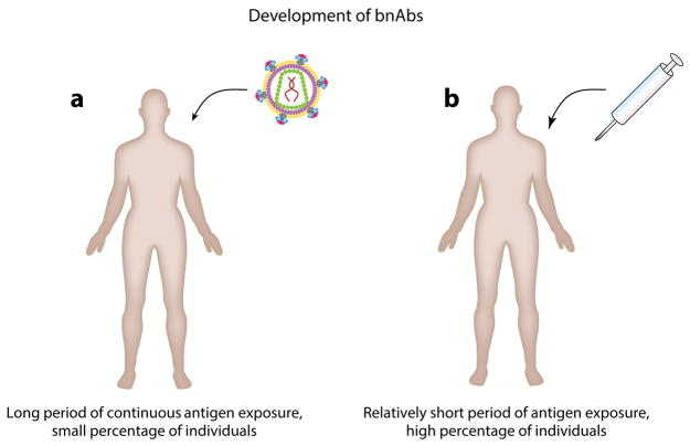
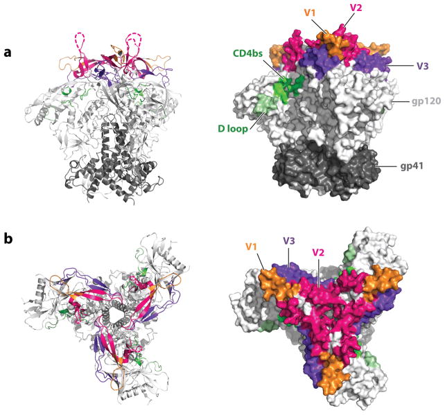
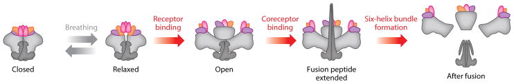
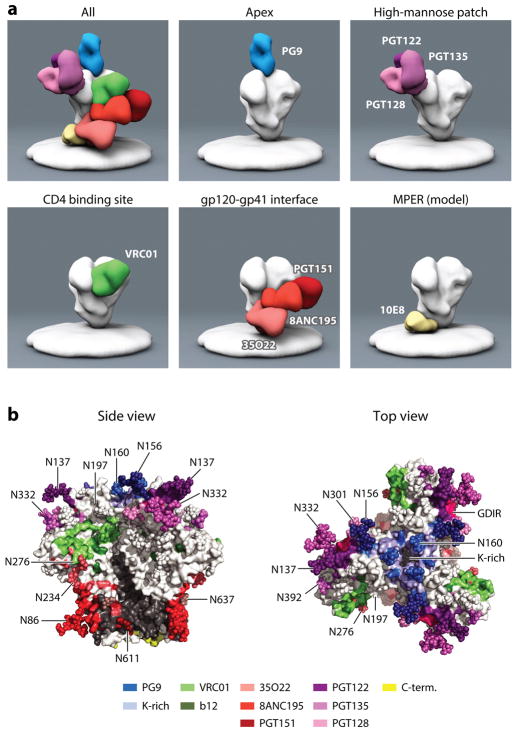
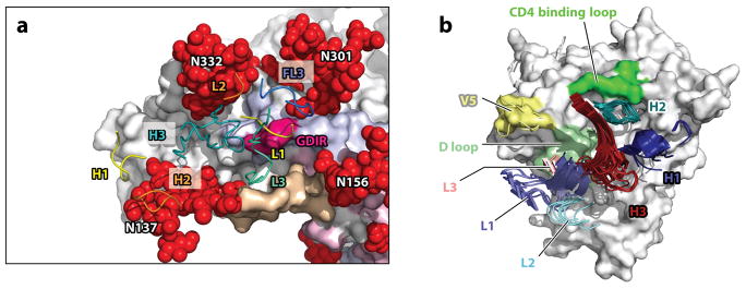
HIV employs multiple means to evade the humoral immune response, particularly the elicitation of and recognition by broadly neutralizing antibodies (bnAbs). Such antibodies can act antivirally against a wide spectrum of viruses by targeting relatively conserved regions on the surface HIV envelope trimer spike. Elicitation of and recognition by bnAbs are hindered by the arrangement of spikes on virions and the relatively difficult access to bnAb epitopes on spikes, including the proximity of variable regions and a high density of glycans. Yet, in a small proportion of HIV-infected individuals, potent bnAb responses do develop, and isolation of the corresponding monoclonal antibodies has been facilitated by identification of favorable donors with potent bnAb sera and by development of improved methods for human antibody generation. Molecular studies of recombinant Env trimers, alone and in interaction with bnAbs, are providing new insights that are fueling the development and testing of promising immunogens aimed at the elicitation of bnAbs.

Keywords: conserved epitopes; immunization strategies; passive immunotherapy; rational vaccine design; vaccination.


Figures

Figure 1
Figure 1
The essence of the problem of bnAbs in HIV vaccine design. (a) Potent bnAb responses develop in a small proportion of HIV-infected individuals, but this occurs over a long period (years) during which the individual is exposed to multiple viral strains. (b) A vaccine should induce such responses in the majority of individuals in a much shorter period with a manageable number of immunogens.
Figure 2
Figure 2
Structure of the HIV envelope spike. In a crystal structure of the HIV strain BG505 expressed as SOSIP.664 near-native trimers [PDB 4zmj (31)], the location of some of the most important Env trimer domains is indicated as viewed (a) from the side or (b) from the top. The gp120 subunits form the blades of a propeller-like structure, whereas the gp41 subunit forms a central stalk and a membrane-proximal, pedestal-like structure. As predicted from other experimental data, hypervariable loops V1 through V3 are located at the apex of the trimer, with hypervariable loop V3 being partially buried under hypervariable loops V1 and V2. These loops are also in proximity to the CD4 receptor binding site that is recessed left in the cleft formed by two propeller blades. Loops that did not resolve in the structure are indicated by a dashed line.
Figure 3
Figure 3
Conformations of the Env trimer. Several conformations have been described for the Env trimer. Before receptor engagement, and depending on the isolate, the Env trimers transit between closed, slightly more open, and relaxed conformations in a process referred to as breathing. Following CD4 receptor engagement, the trimers open out and the CCR5 (or CXCR4) coreceptor binding sites become available. Coreceptor binding then triggers insertion of the fusion peptide into the host membrane and formation of the six-helix bundle that pulls the host and the virus membrane into fusion proximity.
Figure 4
Figure 4
Breadth and potency of bnAbs. The percentage of large panels of isolates neutralized at an IC50 < 50 μg/mL (coverage) in pseudovirus assays plotted against the median inhibitory concentration (IC50 in μg/mL) for all neutralization-sensitive viruses. First-generation antibodies are indicated by unfilled circles and second-generation antibodies by filled circles. The epitope specificity is indicated by color, with antibody specific for the CD4 binding site depicted in green, apex-specific antibodies in blue, antibodies binding to the high-mannose patch in purple, antibodies binding to the gp120-gp41 interface in red, and MPER-specific antibodies in light brown. Note that different panels of viruses have been used for many of the antibodies, so comparisons are approximate (–, , –86, 159). The locations of the epitopes recognized by the bnAbs are illustrated in Figure 5.
Figure 5
Figure 5
(a) Negative-stain electron microscopy representations of bnAbs binding to the Env trimer. bnAbs can be divided according to the epitopes they recognize: those specific for the CD4 binding site, the apex, the high-mannose patch, the gp120-gp41 interface, and the MPER. Electron microscopy reconstructions, with the exception of the MPER panel, were kindly prepared by Jeong Hyun Lee, Gabriel Ozorowski, and Andrew Ward. The MPER antibody reconstruction has been very roughly modeled according to the location of the MPER epitope in the Env trimer structures. (b) The footprints of bnAbs on the Env trimer. The Env trimer coordinates from PDB 4TVP were extracted and stained in white and gray for gp120 and gp41. The contact residues for PG9 are blue (99); PGT122 dark purple (22); PGT128 light purple (96); PGT135 medium purple (136); VRC01 green (88); b12 olive green, from alanine scan (160); PGT151 dark red (98); 8ANC195 medium red (86, 87); and 35O22 light red (24). The MPER region is largely missing from the structure; the last available amino acids are in yellow. The glycan at position 156 of gp120 is recognized by both PGT9 and PGT122, which is reflected by the intermediate color. The glycan at position 332 is bound by most bnAbs specific for the high-mannose patch. Also, the epitopes of PGT151 and 8ANC195 overlap in recognition of the glycan at position 637. The lysine-rich region at the apex is indicated by bright blue. Note that glycans in the structure might be truncated given that they were not completely resolved in the crystal structure.
Figure 6
Figure 6
Recognition of Env by bnAbs. (a) Interaction of PGT122 with the high-mannose patch epitope of BG505 SOSIP [PDB 4NCO (22)]. Only CDR1 through 3 of the heavy and light antibody chains are shown, in yellow, orange, and teal, respectively. gp120 is shown in white, gp41 in gray. Glycans are depicted as red spheres, and gp120 hypervariable regions 1 through 2 are shown in tan, pink, and light blue, respectively. Close contact with multiple glycans is illustrated. The protein surface contacted is the GDIR amino acid sequence at the base of the V3 hypervariable loop. (b) Superimposition of the complementarity-determining regions of multiple VRC01-like antibodies bound to gp120 (PDB 4jpv, 4jpw, 4lsp, 4lsq, 4lsr, 4lss, 4lst, 4lsu, and 4lsv adapted from Reference 89). CDR L1 and H1 are indicated in light and dark blue, CDR L2 and H3 in light and dark teal, CDR L3 and H3 in light and dark red. The CD4 binding loop is in bright green, the D loop in pale green, and the V5 loop in yellow.

References

    1. Corti D, Lanzavecchia A. Broadly neutralizing antiviral antibodies. Annu Rev Immunol. 2013;31(1):705–42. - PubMed
    1. Burton DR, Poignard P, Stanfield RL, Wilson IA. Broadly neutralizing antibodies present new prospects to counter highly antigenically diverse viruses. Science. 2012;337(6091):183–86. - PMC - PubMed
    1. Zanetti G, Briggs JA, Grunewald K, Sattentau QJ, Fuller SD. Cryo-electron tomographic structure of an immunodeficiency virus envelope complex in situ. PLOS Pathog. 2006;2(8):e83. - PMC - PubMed
    1. Zhu P, Liu J, Bess J, Chertova E, Lifson JD, et al. Distribution and three-dimensional structure of AIDS virus envelope spikes. Nature. 2006;441(7095):847–52. - PubMed
    1. Brandenberg OF, Magnus C, Rusert P, Regoes RR, Trkola A. Different infectivity of HIV-1 strains is linked to number of envelope trimers required for entry. PLOS Pathog. 2015;11(1):e1004595. - PMC - PubMed

Publication types