. 2016 Sep;229(3):459-72.
doi: 10.1111/joa.12483. Epub 2016 May 11.

Uterine morphology during diapause and early pregnancy in the tammar wallaby (Macropus eugenii)

Affiliations

Uterine morphology during diapause and early pregnancy in the tammar wallaby (Macropus eugenii)

Melanie K Laird et al. J Anat. 2016 Sep.

Abstract

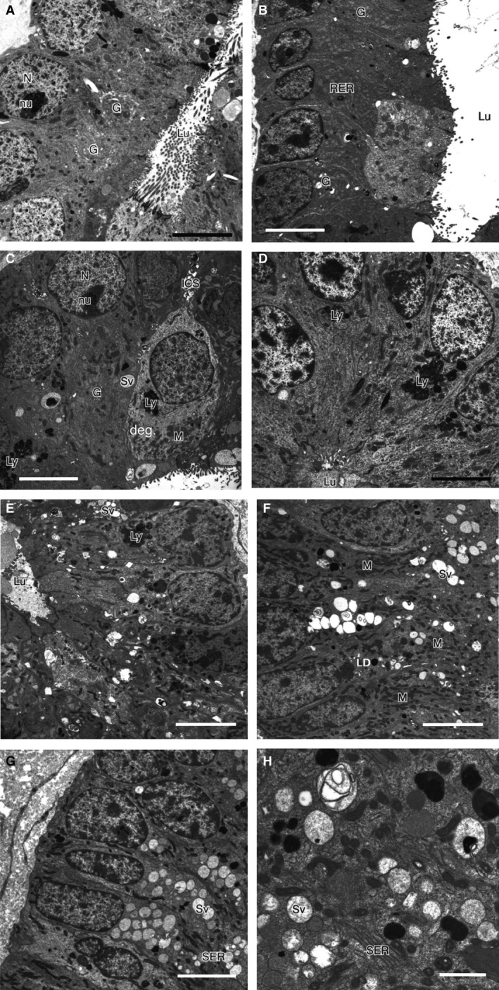
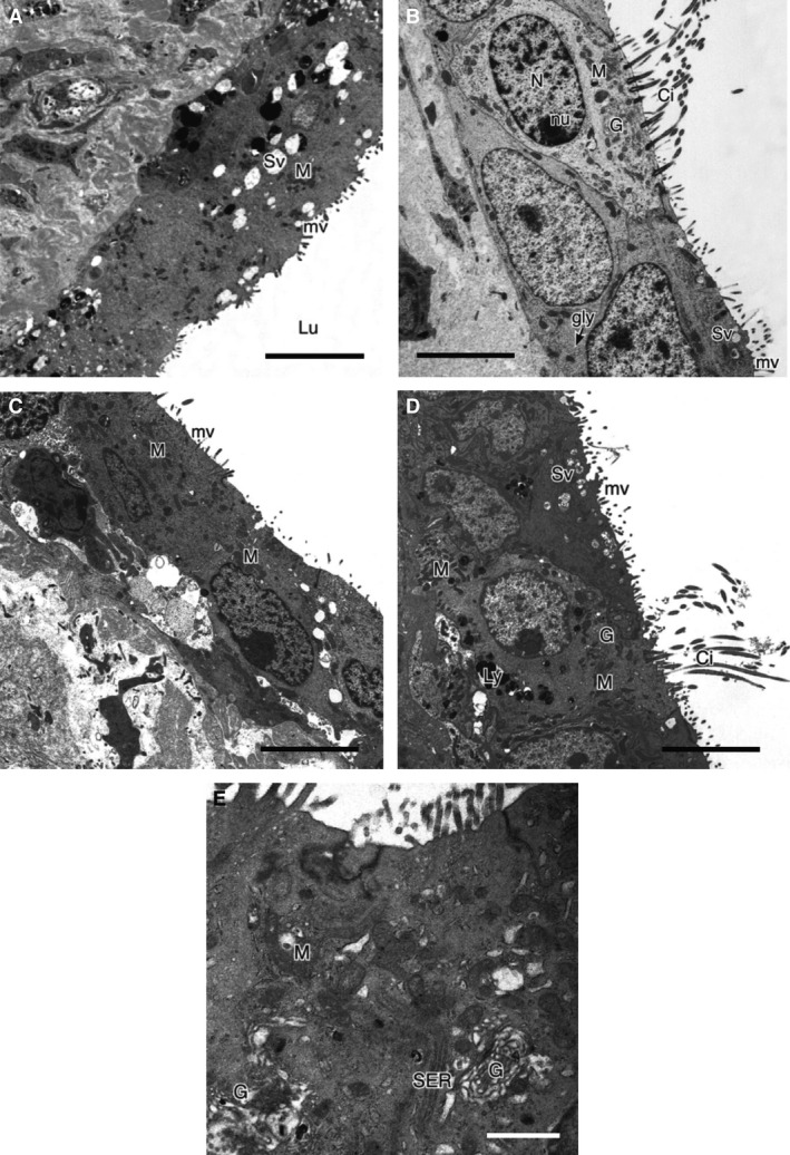
In mammals, embryonic diapause, or suspension of embryonic development, occurs when embryos at the blastocyst stage are arrested in growth and metabolism. In the tammar wallaby (Macropus eugenii), there are two separate uteri, only one of which becomes gravid with the single conceptus at a post-partum oestrus, so changes during pregnancy can be compared between the gravid and non-gravid uterus within the same individual. Maintenance of the viable blastocyst and inhibition of further conceptus growth during diapause in the tammar is completely dependent on the uterine environment. Although the specific endocrine and seasonal signals are well established, much less is known about the cellular changes required to create this environment. Here we present the first detailed study of uterine morphology during diapause and early pregnancy of the tammar wallaby. We combined transmission electron microscopy and light microscopy to describe the histological and ultrastructural changes to luminal and glandular epithelial cells. At entry into diapause after the post-partum oestrus and formation of the new conceptus, there was an increase in abundance of organelles associated with respiration in the endometrial cells of the newly gravid uterus, particularly in the endoplasmic reticulum and mitochondria, as well as an increase in secretory activity. Organelle changes and active secretion then ceased in these cells as they became quiescent and remained so for the duration of diapause. In contrast, cells of the non-gravid, post-partum, contralateral uterus underwent sloughing and remodelling during this time and some organelle changes in glandular epithelial cells continued throughout diapause, suggesting these cells are not completely quiescent during diapause, although no active secretion occurred. These findings demonstrate that diapause, like pregnancy, is under unilateral endocrine control in the tammar, and that preparation for and maintenance of diapause requires substantial changes to uterine endometrial cell ultrastructure and activity.

Keywords: diapause; endometrium; tammar; transmission electron microscopy; uterus.


Figures

Figure 1
Figure 1
Reproductive cycle of the tammar wallaby (Macropus eugenii). The uterus associated with the foregone pregnancy is termed the post‐partum uterus. The contralateral uterus which receives the embryo, and is associated first with the Graafian follicle, then with the new corpus luteum (CL) following ovulation, is termed the newly gravid uterus.
Figure 2
Figure 2
Summary of the key uterine changes during pregnancy and diapause in the tammar wallaby (Macropus eugenii) associated with changes in the ovary and embryo; CL, Corpus luteum; G, Golgi complex; G follicle, Graafian follicle; GC, granulosa cells; GE, glandular epithelium; LC, luteal cells; LE, luminal epithelium of uterus; M, mitochondria; RER, rough endoplasmic reticulum; SER, smooth endoplasmic reticulum; sp, sperm; SVes, secretory vesicles. 1Rudd (1994); 2Renfree & Lewis (1996), Tyndale‐Biscoe (1986), Renfree (1993); 3Renfree et al. (2011).
Figure 3
Figure 3
Histological sections of the newly gravid and post‐partum uteri of the tammar wallaby. The uterus is lined by the luminal epithelium (le), with uterine glands (gl) embedded in the connective tissue stroma (st), which is underlaid by myometrium (m). Sections are stained with haemotoxylin and eosin. (A‐E) Newly gravid uterus: (A) day 0 post‐partum; (B) day 1 post‐partum; (C) day 5 post‐partum; (D) day 8 post‐partum; (E) diapause. (F–J) Post‐partum uterus: (F) day 0 post‐partum; (G) day 1 post‐partum; (H) day 5 post‐partum; (I) day 8 post‐partum; (J) diapause; Scale bar: 100 μm.
Figure 4
Figure 4
Measurements of uterine tissues in the tammar wallaby from days 0–8 post‐partum and in diapause (early, middle, late). (A) Abundance of endometrial gland profiles (cross‐sections) per 0.5 mm2. (B) Thickness of the luminal epithelium. (C) Thickness of the endometrial stroma. Measurements (means ± SEM) are from both post‐partum (open symbols) and newly gravid uteri (solid symbols) from day 0 to day 8 post‐partum, and during embryonic diapause. Asterisks denote values that are significantly different from the day of parturition (day 0 post‐partum), where *P < 0.05; **P < 0.01. A timeline shows the stage of embryonic development relative to the time of birth: ovulation (Ov); embryo enters the uterus (Ut); cleavage stage of the embryo in the uterus (2, 8, 16, 32 or 64 cells); formation of the blastocyst in the days after birth.
Figure 5
Figure 5
Transmission electron micrographs of glandular epithelial cells of the newly gravid endometrium. Scale bar: 5 μm. (A) Day 0 post‐partum. (B) Day 1–2 post‐partum. (C) Day 3–5 post‐partum. (D) Day 6–8 post‐partum. (E) Diapause. (F) Higher magnification of (E). Scale bar: 1 μm. bm, basement membrane; G, Golgi complex, L, lipid droplets; Lu, glandular lumen; Ly, lysosomes; M, mitochondria; mv, microvilli, N, nucleus; RER, rough endoplasmic reticulum; s, secretory material; SER, smooth endoplasmic reticulum; SV, secretory vesicles.
Figure 6
Figure 6
Transmission electron micrographs of glandular epithelial cells of the post‐partum endometrium. Scale bar: 5 μm. (A) Day 0 post‐partum. (B) Day 1–2 post‐partum. (C) Day 3 post‐partum. (D) Day 5 post‐partum. deg, degenerative cells; G, Golgi complex; ICS, intercellular space, Lu, glandular lumen; Ly, lysosomes; M, mitochondria; N, nuclei; RER, rough endoplasmic reticulum; Sv, secretory vesicles. (E) Day 6–8 post‐partum. (F) Mid diapause. (G) Late diapause. (H) Higher magnification of (G). Scale bar: 1 μm. LD, lipid droplets; Lu, glandular lumen; Ly, lysosomes; M, elongated mitochondria; SER, smooth endoplasmic reticulum; Sv, secretory vesicles.
Figure 7
Figure 7
Transmission electron micrographs of luminal epithelial cells of the newly gravid endometrium. Scale bar: 4 μm. (A) Day 0 post‐partum. (B) Day 1 post‐partum. (C) Day 3–5 post‐partum. (D) Day 6–8 post‐partum. (E) Diapause. Scale bar:  1 μm. bm, basement membrane; Ci, cilia; G, Golgi complex; gly, glycogen granules; gr, dark granules; L, lipid droplets; Lu, uterine lumen; Ly, lysosomes; M, basal mitochondria; mv, microvilli; N, nuclei; SER, smooth endoplasmic reticulum; Sv, secretory vesicles; (→) active exocytosis of secretory granules.
Figure 8
Figure 8
Transmission electron micrographs of luminal epithelial cells of the post‐partum endometrium. Scale bar:  4 μm. (A) Day 0 post‐partum. (B) Day 1 post‐partum. (C) Day 3–5 post‐partum. (D) Day 6–8 post‐partum. (E) Diapause. Scale bar:  1 μm. Ci, cilia; G, Golgi complex; gly, glycogen granules; Lu, uterine lumen; Ly, lysosomes; M, basal mitochondria; mv, microvilli; N, nuclei; SER, smooth endoplasmic reticulum; Sv, secretory vesicles.

References

    1. Cha J, Sun X, Bartos A, et al. (2013) A new role for muscle segment homeobox genes in mammalian embryonic diapause. Open Biol 3, 130035. - PMC - PubMed
    1. Cruz YP, Selwood L (1993) Uterine histology of the dasyurid marsupial, Antechinus stuartii: relationship with differentiation of the embryo. J Reprod Fertil 99, 237–242. - PubMed
    1. Cruz YP, Selwood L (1997) Histological differences between gravid and non‐gravid uteri in the dasyurid marsupial, Sminthopsis macroura . J Reprod Fertil 111, 319–325. - PubMed
    1. Curlewis JD, Stone GM (1986) Effects of oestradiol, the oestrous cycle and pregnancy on weight, metabolism and cytosol receptors in the uterus of the brush‐tail possum (Trichosurus vulpecula). J Endocrinol 108, 201–210. - PubMed
    1. Daniel JC (1971) Growth of the preimplantation embryo of the northern fur seal and its correlation with changes in uterine protein. Dev Biol 26, 316–322. - PubMed

Publication types

LinkOut - more resources