Sclerosing hemangioma: A diagnostic dilemma in fine needle aspiration cytology
- PMID: 27168758
- PMCID: PMC4854032
- DOI: 10.4103/1742-6413.180783
Sclerosing hemangioma: A diagnostic dilemma in fine needle aspiration cytology
Abstract
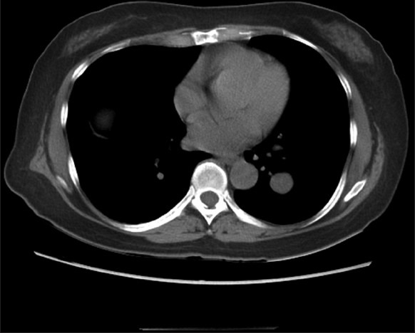
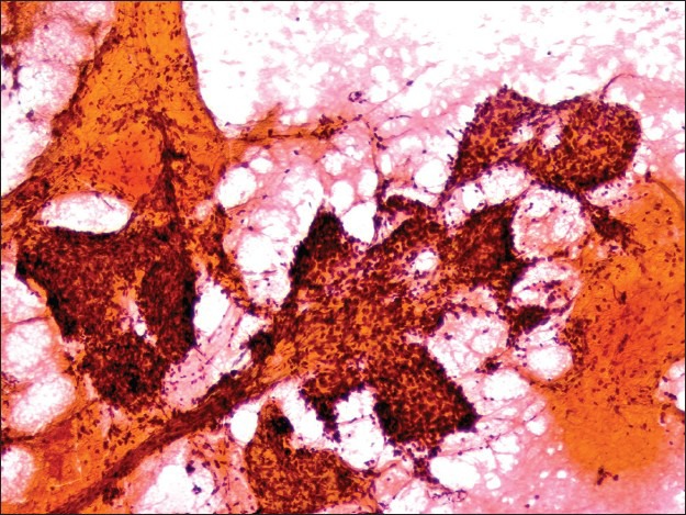
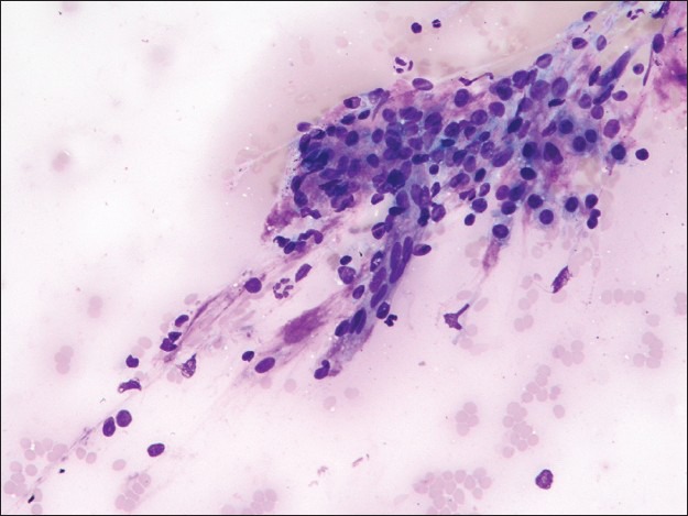
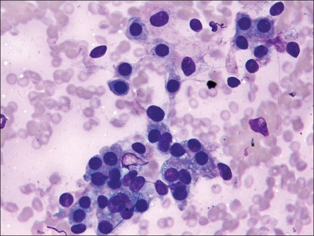
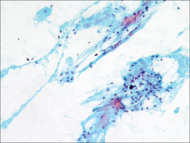
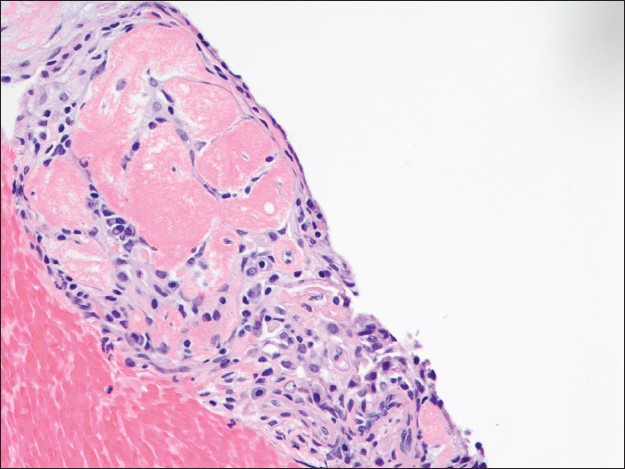
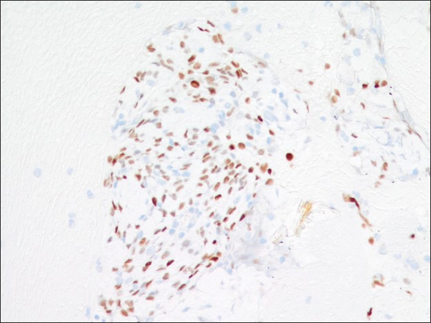
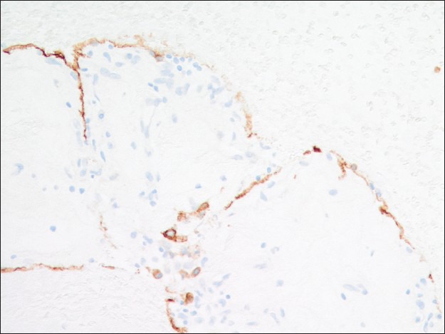
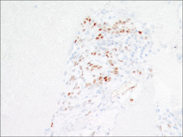
Sclerosing hemangioma of the lung is a benign neoplasm with a widely debated histogenesis. It has a polymorphic histomorphology characterized by a biphasic cell population of "surface cells" and "round cells" arranged in four general patterns: Papillary, solid, angiomatous, and sclerotic. This variability in histomorphology makes it difficult to diagnose sclerosing hemangioma by fine needle aspiration (FNA). We present a case of sclerosing hemangioma diagnosed on FNA with immunohistochemistry performed on an accompanied cell block. The clinical presentation, cytomorphology, immunohistochemistry, and differential diagnoses are discussed.
Keywords: Cytology; fine needle aspiration; lung; pneumocytoma; sclerosing hemangioma.
Figures
References
-
- Liebow AA, Hubbell DS. Sclerosing hemangioma (histiocytoma, xanthoma) of the lung. Cancer. 1956;9:53–75. - PubMed
-
- Wojcik EM, Sneige N, Lawrence DD, Ordóñez NG. Fine-needle aspiration cytology of sclerosing hemangioma of the lung: Case report with immunohistochemical study. Diagn Cytopathol. 1993;9:304–9. - PubMed
-
- Fukayama M, Koike M. So-called sclerosing hemangioma of the lung. An immunohistochemical, histochemical and ultrastructural study. Acta Pathol Jpn. 1988;38:627–42. - PubMed
Publication types
LinkOut - more resources
Full Text Sources
Other Literature Sources