. 2016 May;11(5):1893-1895.
doi: 10.3892/etm.2016.3097. Epub 2016 Feb 19.

Local recurrent vaginal aggressive angiomyxoma misdiagnosed as cellular angiomyofibroblastoma: A case report

Affiliations

Local recurrent vaginal aggressive angiomyxoma misdiagnosed as cellular angiomyofibroblastoma: A case report

Yin-Feng Wang et al. Exp Ther Med. 2016 May.

Abstract

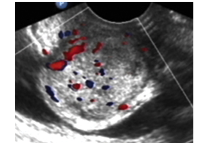
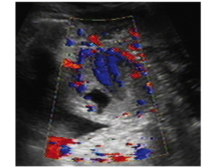
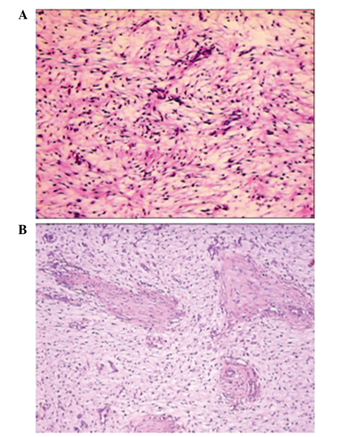
Aggressive angiomyxoma (AAM) and angiomyofibroblastoma (AMFB) are two rare types of mesenchymal tumors with overlapping clinicopathological features. In certain cases, the differential diagnosis between the two tumors is difficult even for experienced pathologists. The present study reported the case of a well-circumscribed soft tissue mass on the anterior wall of the vagina in a 25-year-old woman. The mass was initially removed without disturbance to the adjacent tissues. The histopathological features included spindle cells in inconspicuous myxoid stroma and a well-demarcated mass without evidence of invasion, which prompted the initial diagnosis of AMFB. After 2 years, a mass returned in the same area and a wide tumor excision was performed. The histopathological examination confirmed the final diagnosis of AAM upon review.

Keywords: aggressive angiomyxoma; cellular angiomyofibroblastoma; misdiagnosis; recurrence.


Figures

Figure 1.
Figure 1.
Transvaginal ultrasound revealed a well-circumscribed soft tissue mass with heterogeneous internal echos. The size of the mass was 3.3×3.7×3.4 cm, with rich intralesional vascularization. The blood flow was investigated using color Doppler.
Figure 2.
Figure 2.
Transvaginal ultrasound demonstrated a hypoechoic circumscribed but not well-demarcated mass. Color Doppler flow imaging revealed predominantly low resistance type arterial blood flow between the paries anterior vagina and posterior bladder wall. The size of the mass was 4.9×3.6×3.0 cm.
Figure 3.
Figure 3.
Histopathological changes indicated similar morphological features in the (A) initial and (B) second surgeries, including cellular spindle cells and small blood vessels in the myxoid stroma. However, the specimen from the second surgery displayed sparser cells and more prominent myxoid stroma, compared with the initial surgery specimen. The small elastic vessels in the two figures is not a typical feature of AAM; they are more frequently observed in AMFB. Magnification, ×100.

References

    1. Steeper TA, Rosai J. Aggressive angiomyxoma of the female pelvis and perineum. Report of nine cases of a distinctive type of gynecologic soft-tissue neoplasm. Am J Surg Pathol. 1983;7:463–475. doi: 10.1097/00000478-198307000-00009. - DOI - PubMed
    1. Bakhtiar UJ, Awan AS. Aggressive angiomyxoma of vulva. J Coll Physicians Surg Pak. 2013;23:507–508. - PubMed
    1. Ki EY, Park JS, Lee A, Hur SY. Aggressive angiomyxoma of the female genital tract: Report of two cases. Eur J Gynaecol Oncol. 2014;35:465–468. - PubMed
    1. Sengupta SK, Bhattacharyya SK, Saha SP, Roy H, Sarkar AN. Recurrent aggressive angiomyxoma of the vulva-a rare presentation. J Clin Diagn Res. 2014;8:OD01–OD02. - PMC - PubMed
    1. Ducarme G, Valentin M, Davitian C, Felce-Dachez M, Luton D. Angiomyofibroblastoma: A rare vulvar tumor. Arch Gynecol Obstet. 2010;281:161–162. doi: 10.1007/s00404-009-1110-4. - DOI - PubMed

LinkOut - more resources