Leg Swelling Caused by Heterotopic Ossification Mimicking Deep Vein Thrombosis in a Paraplegic Patient
- PMID: 27169085
- PMCID: PMC4847498
- DOI: 10.13004/kjnt.2015.11.2.158
Leg Swelling Caused by Heterotopic Ossification Mimicking Deep Vein Thrombosis in a Paraplegic Patient
Abstract
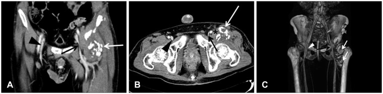
Leg swelling in patients with paraplegia due to spinal cord injury (SCI) occurs for various reasons, including heterotopic ossification (HO), deep vein thrombosis (DVT), fracture, or cellulitis. The clinical presentations of these conditions may overlap in part or in whole and it may occasionally be difficult to distinguish. Of these conditions, DVT and subsequent pulmonary embolism remain significant causes of morbidity and mortality in patients with SCI. Therefore, a prompt diagnostic work-up, particularly for DVT, is essential in patients with SCI, who present with leg swelling. Here, we report a case of leg swelling in a paraplegic patient, resulting from HO mimicking DVT and discuss the differential diagnosis.
Keywords: Ossification, heterotopic; Paraplegia; Spinal cord injuries; Venous thrombosis.
Conflict of interest statement
The authors have no financial conflicts of interest.
Figures
References
-
- Bekou V, Galis D, Traber J. Unilateral leg swelling: deep vein thrombosis? Phlebology. 2011;26:8–13. - PubMed
-
- Blankenship LD, Strommen JA. 27-year-old man with a swollen leg. Mayo Clin Proc. 2000;75:977–980. - PubMed
-
- Citak M, Suero EM, Backhaus M, Aach M, Godry H, Meindl R, et al. Risk factors for heterotopic ossification in patients with spinal cord injury: a case-control study of 264 patients. Spine (Phila Pa 1976) 2013;37:1953–1957. - PubMed
Publication types
LinkOut - more resources
Full Text Sources
Other Literature Sources