. 2016 May 11;9(1):53.
doi: 10.1186/s13041-016-0234-2.

LRRK2 phosphorylation level correlates with abnormal motor behaviour in an experimental model of levodopa-induced dyskinesias

Affiliations

LRRK2 phosphorylation level correlates with abnormal motor behaviour in an experimental model of levodopa-induced dyskinesias

Jennifer Stanic et al. Mol Brain. .

Abstract

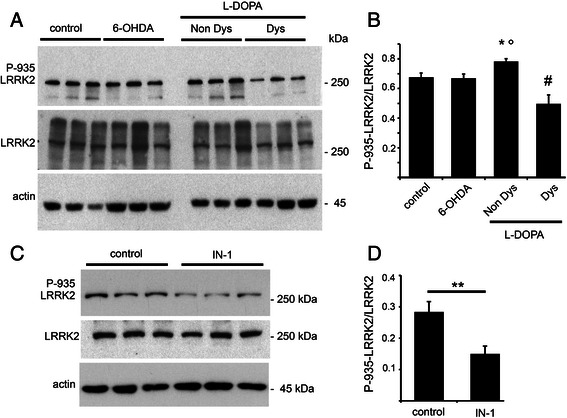
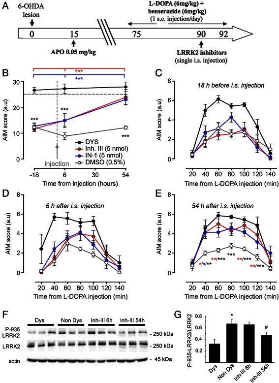
Levodopa (L-DOPA)-induced dyskinesias (LIDs) represent the major side effect in Parkinson's disease (PD) therapy. Leucine-rich repeat kinase 2 (LRRK2) mutations account for up to 13 % of familial cases of PD. LRRK2 N-terminal domain encompasses several serine residues that undergo phosphorylation influencing LRRK2 function. This work aims at investigating whether LRRK2 phosphorylation/function may be involved in the molecular pathways downstream D1 dopamine receptor leading to LIDs. Here we show that LRRK2 phosphorylation level at serine 935 correlates with LIDs induction and that inhibition of LRRK2 induces a significant increase in the dyskinetic score in L-DOPA treated parkinsonian animals. Our findings support a close link between LRKK2 functional state and L-DOPA-induced abnormal motor behaviour and highlight that LRRK2 phosphorylation level may be implicated in LIDs, calling for novel therapeutic strategies.

Keywords: 6-OHDA; L-DOPA; L-DOPA-induced dyskinesias; LRRK2; Parkinson’s disease; Phosphorylation; Rat.


Figures

Fig. 1
Fig. 1
LRRK2 phosphorylation at Ser-935 in parkinsonian and dyskinetic rats. a The ipsilateral striatum from non lesioned (control), fully lesioned parkinsonian (6-OHDA), non dyskinetic (Non Dys) rats or animals displaying a dyskinetic behaviour (Dys) were analyzed by WB to evaluate the levels of LRRK2 phosphorylation at Ser-935 site (P-935). b The graph illustrates LRRK2 phosphorylation normalized upon total LRRK2 (n = 6; One-way Anova: *p < 0.05 Non Dys versus 6-OHDA °p < 0.01 Non dys versus Dysk, #p < 0.05, Dys versus 6-OHDA). Data are expressed as mean ± SEM. c Rats were injected in the left striatum with LRRK2 IN-1 inhibitor or vehicle (control) and the total and phosphorylated LRRK2 levels were evaluated by WB. d IN-1 reduced LRRK2 phosphorylation in vivo (n = 6; **p < 0.01, unpaired two-tailed Student’s t-test). Graph reports LRRK2 phosphorylation level normalized on the total amount of LRRK2 protein. Data are expressed as mean ± SEM
Fig. 2
Fig. 2
Effects of LRRK2 inhibitors on dyskinesia in 6-OHDA parkinsonian rats. a The panel is a schematic representation of the experimental design. Rats were unilaterally injected with 6-OHDA in the left MFB. After 15 days, the extent of the lesion was evaluated upon apomorphine answer. Two months after the 6-OHDA injection, fully lesioned rats (>200 controlateral turns) were chronically treated with L-DOPA. LRRK2 inhibitors were administrated to low dyskinetic animals with a single intrastriatal (i.s.) injection after 2 weeks of L-DOPA treatment. LIDs onset and severity were evaluated by AIMs scoring. b IN-1 (5 nmol), LRRK2 inhibitor III (5 nmol) or vehicle (5 μl 0.5 % DMSO) were stereotaxically injected in the ipsilateral striatum of low dyskinetic rats. I.s. injections were performed 6 h before the daily L-DOPA administration and the evaluation of AIMs were carried out from 20 to 140 min after L-DOPA. Both inhibitors were able to induce a significant increase in the AIMs score compared to vehicle injected rats and to pre-surgery values (n = 7; −18 h: ***p < 0.001, DYS vs DMSO/IN-1/Inh. III. 6 h: ***p < 0.001, DYS vs DMSO/IN-1/Inh. III. 54 h: ***p < 0.001, DMSO vs IN-1/Inh. III/DYS. IN-1: ***p < 0.001, 54 h vs −18 h/6 h. Inhibitor III: ***p < 0.001, 54 h vs −18 h/6 h vs 54 h; two-way ANOVA followed by Bonferroni post-hoc test). c-e Both inhibitors significantly increased dyskinesia induction, as indicated by AIMs scoring during the single last observation session (54 h; E) (n = 7; 40 min, DMSO vs IN-1/Inh. III: *p < 0.05, DMSO vs DYS: **p < 0.01; 60 min, DMSO vs Inh. III: **p < 0.01, DMSO vs DYS: ***p < 0.001; 80 min, DMSO vs DYS: ***p < 0.001; 100 min, DMSO vs Inh. III: **p < 0.01, DMSO vs DYS: ***p < 0.001; 120 min, DMSO vs Inh. III: **p < 0.01, DMSO vs DYS: ***p < 0.001; two-way ANOVA plus Bonferroni post-hoc test). f The ipsilateral striatum from rats displaying a dyskinetic behaviour (Dys), non dyskinetic rats (Non Dys), Non Dys animals sacrificed 6 h after Inh-III injection (Inh-III 6 h) or Non Dys animals sacrificed 54 h after Inh-III injection (Inh-III 54 h) were analyzed by WB to evaluate the levels of LRRK2 phosphorylation at Ser-935 site (P-935). g The graph illustrates LRRK2 phosphorylation normalized upon total LRRK2 (n = 3; One-way Anova: *p < 0.01 Non Dys versus Dys #p < 0.05 Non Dys versus Inh-III 54 h). Data are expressed as mean ± SEM

Similar articles

Cited by

References

    1. Bastide MF, Meissner WG, Picconi B, Fasano S, Fernagut P-O, Feyder M, et al. Pathophysiology of L-dopa-induced motor and non-motor complications in Parkinson’s disease. Prog Neurobiol. 2015;132:96–168. doi: 10.1016/j.pneurobio.2015.07.002. - DOI - PubMed
    1. Feyder M, Bonito-Oliva A, Fisone G. L-DOPA-induced dyskinesia and abnormal signaling in striatal medium spiny neurons: focus on dopamine D1 receptor-mediated transmission. Front Behav Neurosci. 2011;5:71. doi: 10.3389/fnbeh.2011.00071. - DOI - PMC - PubMed
    1. Santini E, Valjent E, Usiello A, Carta M, Borgkvist A, Girault J-A, et al. Critical involvement of cAMP/DARPP-32 and extracellular signal-regulated protein kinase signaling in L-DOPA-induced dyskinesia. J Neurosci. 2007;27:6995–7005. doi: 10.1523/JNEUROSCI.0852-07.2007. - DOI - PMC - PubMed
    1. Paisán-Ruíz C, Paisán-Ruíz C, Jain S, Evans EW, Gilks WP, Simón J, et al. Cloning of the gene containing mutations that cause PARK8-linked Parkinson’s disease. Neuron. 2004;44:595–600. doi: 10.1016/j.neuron.2004.10.023. - DOI - PubMed
    1. Zimprich A, Biskup S, Leitner P, Lichtner P, Farrer M, Lincoln S, et al. Mutations in LRRK2 cause autosomal-dominant parkinsonism with pleomorphic pathology. Neuron. 2004;44:601–7. doi: 10.1016/j.neuron.2004.11.005. - DOI - PubMed

MeSH terms