Review
. 2016 Jul;29(3):553-80.
doi: 10.1128/CMR.00076-15.

Dientamoeba fragilis, the Neglected Trichomonad of the Human Bowel

Affiliations
Review

Dientamoeba fragilis, the Neglected Trichomonad of the Human Bowel

Damien Stark et al. Clin Microbiol Rev. 2016 Jul.

Abstract

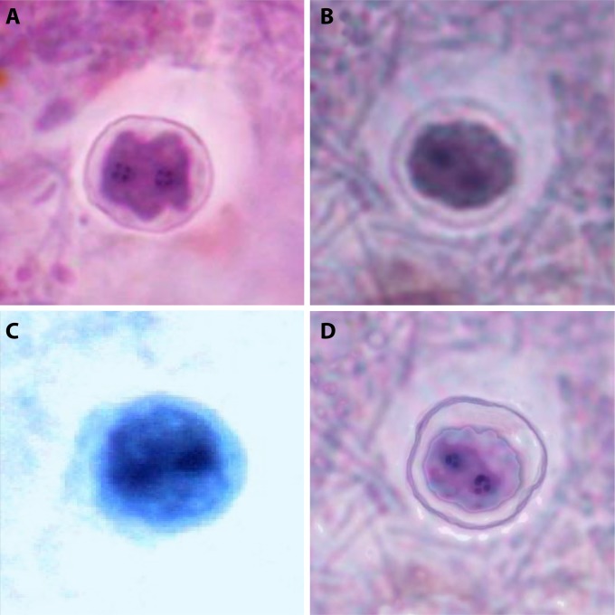
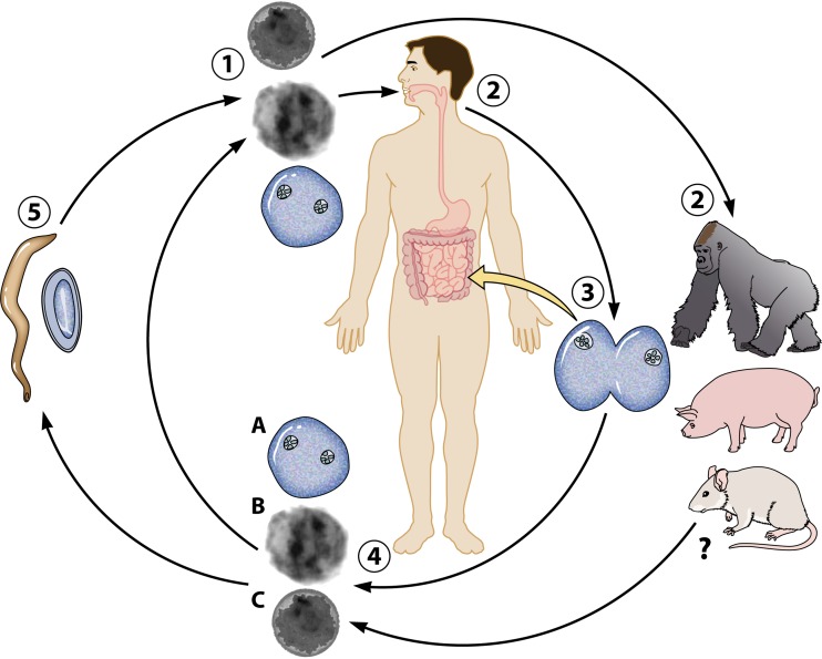
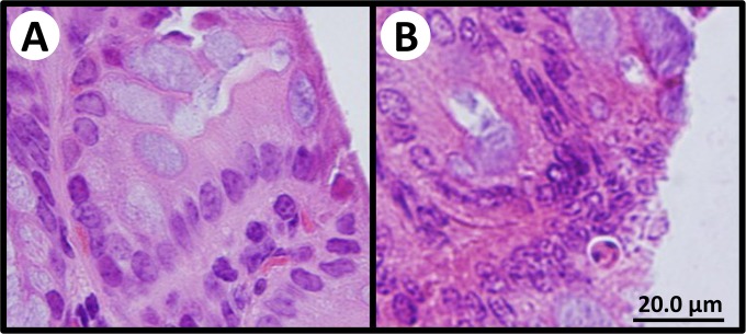
Dientamoeba fragilis is a protozoan parasite of the human bowel, commonly reported throughout the world in association with gastrointestinal symptoms. Despite its initial discovery over 100 years ago, arguably, we know less about this peculiar organism than any other pathogenic or potentially pathogenic protozoan that infects humans. The details of its life cycle and mode of transmission are not completely known, and its potential as a human pathogen is debated within the scientific community. Recently, several major advances have been made with respect to this organism's life cycle and molecular biology. While many questions remain unanswered, these and other recent advances have given rise to some intriguing new leads, which will pave the way for future research. This review encompasses a large body of knowledge generated on various aspects of D. fragilis over the last century, together with an update on the most recent developments. This includes an update on the latest diagnostic techniques and treatments, the clinical aspects of dientamoebiasis, the development of an animal model, the description of a D. fragilis cyst stage, and the sequencing of the first D. fragilis transcriptome.


Figures

FIG 1
FIG 1
Molecular phylogenetic analysis of RNA polymerase II largest-subunit protein sequences from various trichomonads, including D. fragilis. The GenBank accession number for each sequence is shown in parentheses. The evolutionary history was inferred by using the maximum likelihood method based on the Jones, Taylor, Thornton (JTT) matrix-based model (199). The percentage of trees in which the associated taxon cluster together is shown next to the branches. Evolutionary analyses were conducted with MEGA6 (200). The robustness of the tree was assessed by using the bootstrap method with 1,000 replicates.
FIG 2
FIG 2
Pleomorphic trophozoites of D. fragilis. (A) Binucleate trophozoite of D. fragilis (stained with a modified iron-hematoxylin stain) (magnification, ×1,000); (B) live D. fragilis trophozoite displaying motility (viewed under phase-contrast microscopy) (magnification, ×400).
FIG 3
FIG 3
Different cell surface structures observed in D. fragilis trophozoites grown in a xenic culture system. (A and C) Smooth (A) and ruffled (C) cell structures observed by scanning electron microscopy. Ruffled cells accounted for up to 90% of the cell population, although time interval experiments showed an increase in the number of smooth cells at 72 h. (B) Cells with a slightly textured surface are also observed and may represent an intermediate between the smooth and ruffled forms.
FIG 4
FIG 4
Precystic forms from human clinical stool samples (stained with a modified iron-hematoxylin stain) (magnification, ×1,000). Precystic forms are typically smaller (3.5 to 5 μm in diameter) and stain more densely than trophozoites.
FIG 5
FIG 5
Transmission electron micrographs of D. fragilis cysts identified in rodent studies of D. fragilis infection. Dientamoeba cysts are 4 to 6 μm in diameter and possess a distinct cyst wall and a clearly visible peritrophic space.
FIG 6
FIG 6
Binucleate cysts of D. fragilis identified in rodent studies (A and B) and from human clinical stool samples (C and D) (stained with a modified iron-hematoxylin stain) (magnification, ×1,000).
FIG 7
FIG 7
Life cycle of D. fragilis showing current hypotheses on transmission. D. fragilis is ingested from the external environment by a host species in one or more possible forms (1). The preferred transmissible form is yet to be determined. Humans are thought to be the preferred host of D. fragilis, although gorillas, pigs, and rodents are also considered natural hosts (2). Once ingested, D. fragilis travels to the large intestine, where it multiplies by binary fission (3). D. fragilis trophozoites (A), precysts (B), and cysts (C) are passed into the environment in the feces (4), where they contaminate food and/or water sources. D. fragilis parasites are then ingested by a new host, completing the cycle. While D. fragilis trophozoites are infectious to laboratory mice, they are noninfectious to larger mammals. The infectivity of precysts and purified cysts is yet to be demonstrated. It has been suggested that D. fragilis may be transmitted in the ova of the human pinworm, Enterobius vermicularis (5). Recent reports have confirmed the presence of D. fragilis DNA within E. vermicularis ova, although it is unknown whether viable and/or transmissible D. fragilis is present in these ova.
FIG 8
FIG 8
Intact, healthy submucosa of the large intestine of negative-control mice (A) compared to mice infected with D. fragilis (B). Infected mice exhibit mild inflammation and damage to the mucosal layer.
None
Cecil Clifford Dobell, to whom this paper is dedicated.
None
None
None
None

References

    1. Jepps MW, Dobell C. 1918. Dientamoeba fragilis n.g., n. sp.: a new intestinal amoeba from man. Parasitology 10:352–367.
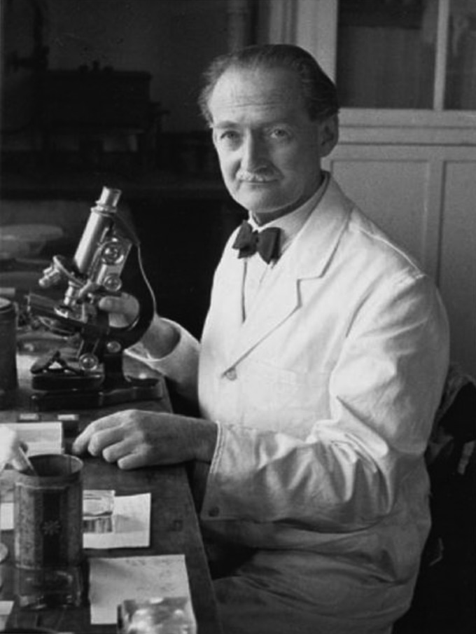
    1. Dobell C. 1940. Researches on intestinal protozoa in monkeys and man. X. The life history of Dientamoeba fragilis: observations, experiments and speculations. Parasitology 32:417–461.
    1. Hess M, Liebhart D, Bilic I, Ganas P. 2015. Histomonas meleagridis—new insights into an old pathogen. Vet Parasitol 208:67–76. doi:10.1016/j.vetpar.2014.12.018. - DOI - PubMed
    1. Ockert G, Schneider W. 1974. Electron microscopic findings in Dientamoeba fragilis. Z Gesamte Hyg 20:555–557. (In German.). - PubMed
    1. Silard R, Burghelea B, Panaitescu D, Burcos V. 1984. Ultrastructure of Dientamoeba fragilis: a study of the mononucleated stage. Arch Roum Pathol Exp Microbiol 43:87–101. - PubMed

Publication types

LinkOut - more resources