. 2016 May 24;113(21):E2983-92.
doi: 10.1073/pnas.1600674113. Epub 2016 May 11.

Restricting nonclassical MHC genes coevolve with TRAV genes used by innate-like T cells in mammals

Affiliations

Restricting nonclassical MHC genes coevolve with TRAV genes used by innate-like T cells in mammals

Pierre Boudinot et al. Proc Natl Acad Sci U S A. .

Abstract

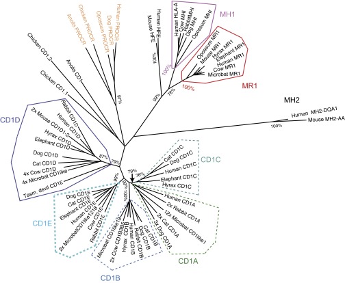
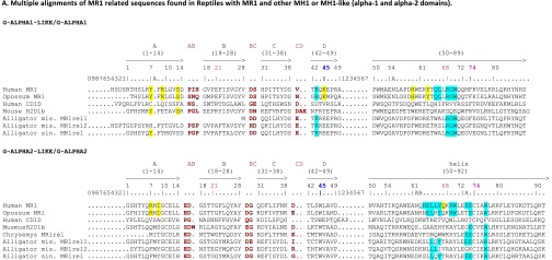
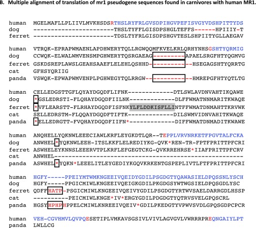
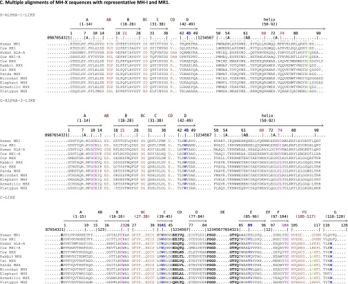
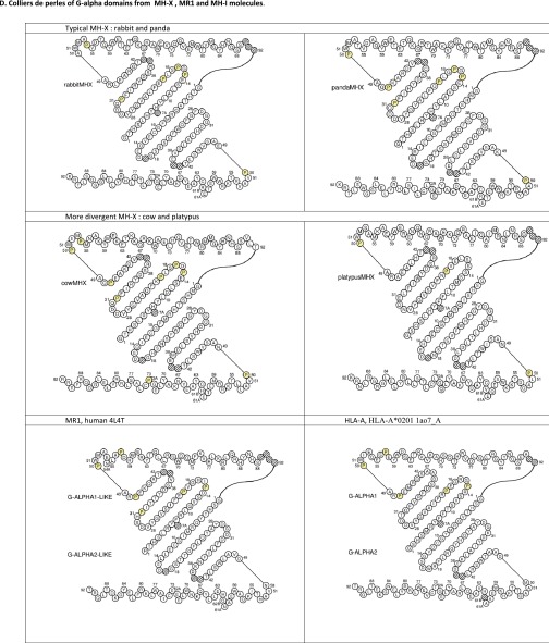
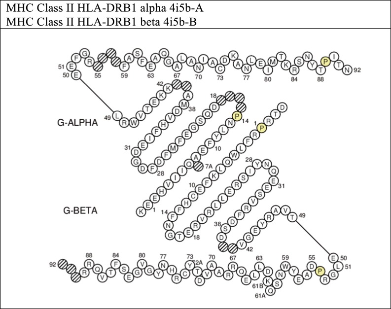
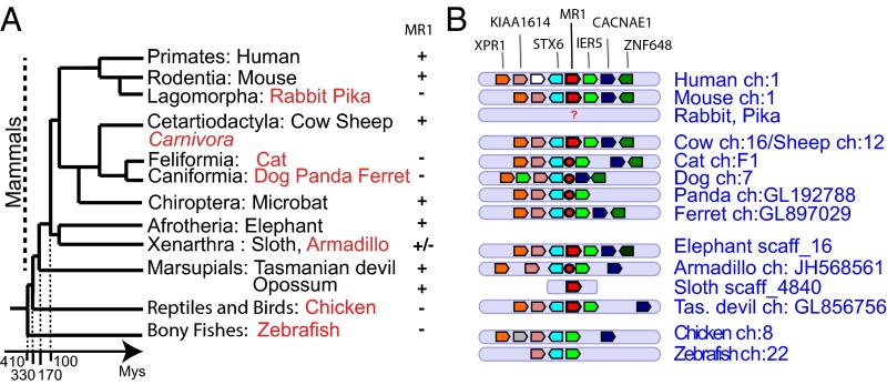
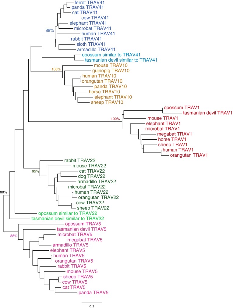
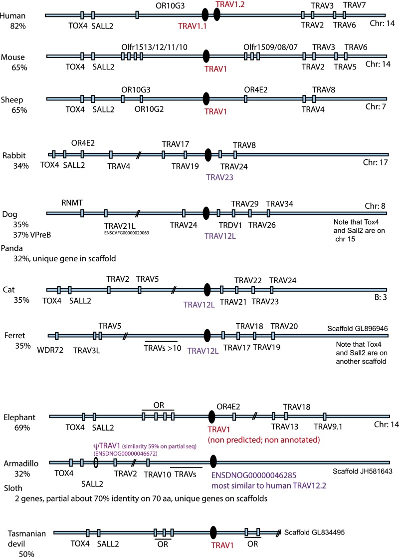
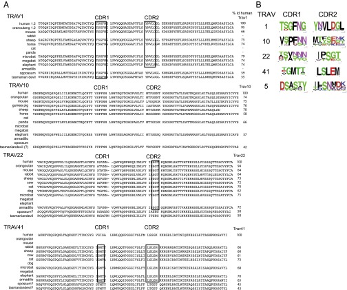
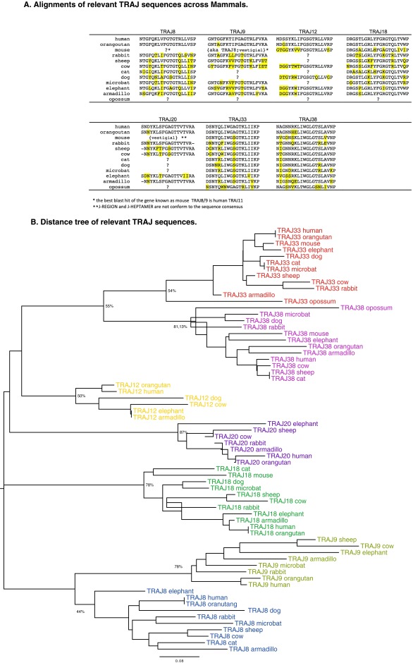
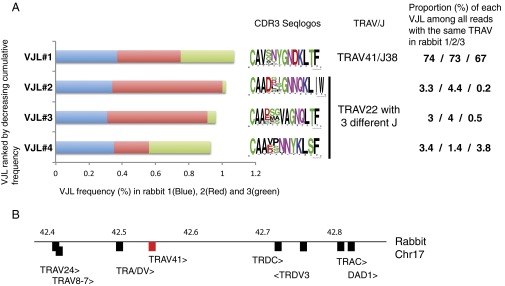
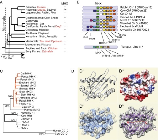
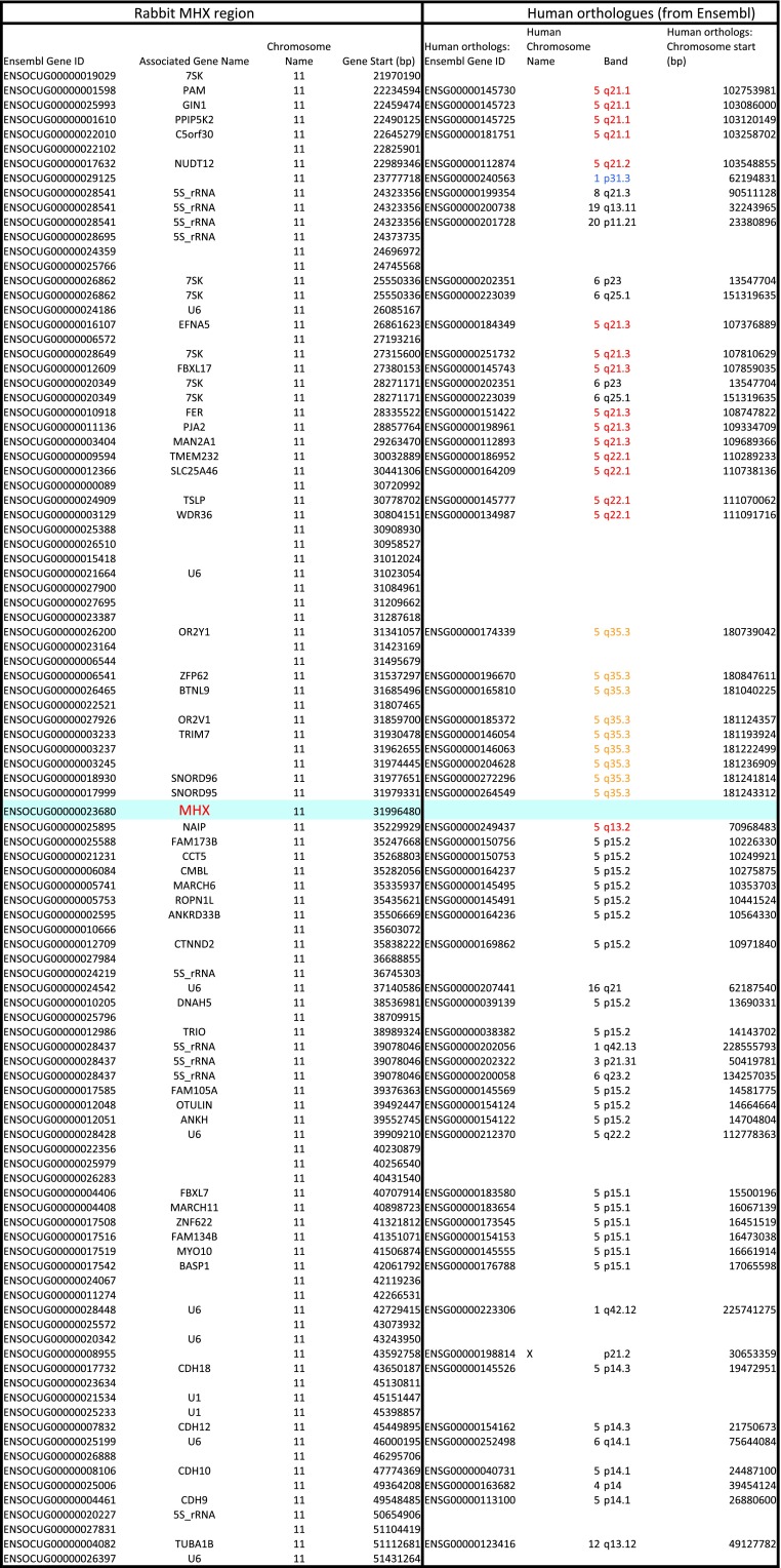
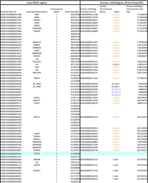
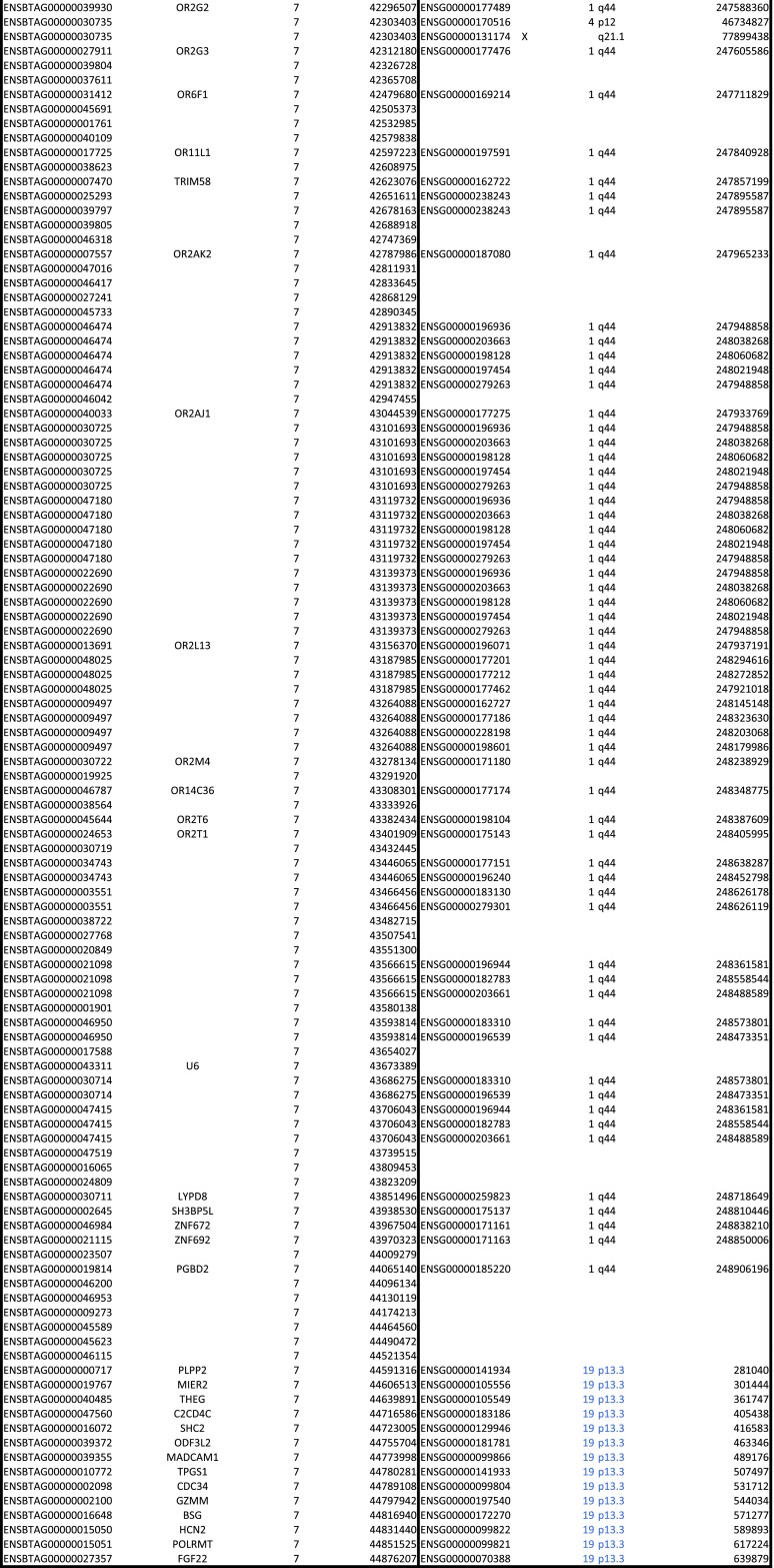
Whereas major histocompatibility class-1 (MH1) proteins present peptides to T cells displaying a large T-cell receptor (TR) repertoire, MH1Like proteins, such as CD1D and MR1, present glycolipids and microbial riboflavin precursor derivatives, respectively, to T cells expressing invariant TR-α (iTRA) chains. The groove of such MH1Like, as well as iTRA chains used by mucosal-associated invariant T (MAIT) and natural killer T (NKT) cells, respectively, may result from a coevolution under particular selection pressures. Herein, we investigated the evolutionary patterns of the iTRA of MAIT and NKT cells and restricting MH1Like proteins: MR1 appeared 170 Mya and is highly conserved across mammals, evolving more slowly than other MH1Like. It has been pseudogenized or independently lost three times in carnivores, the armadillo, and lagomorphs. The corresponding TRAV1 gene also evolved slowly and harbors highly conserved complementarity determining regions 1 and 2. TRAV1 is absent exclusively from species in which MR1 is lacking, suggesting that its loss released the purifying selection on MR1. In the rabbit, which has very few NKT and no MAIT cells, a previously unrecognized iTRA was identified by sequencing leukocyte RNA. This iTRA uses TRAV41, which is highly conserved across several groups of mammals. A rabbit MH1Like gene was found that appeared with mammals and is highly conserved. It was independently lost in a few groups in which MR1 is present, like primates and Muridae, illustrating compensatory emergences of new MH1Like/Invariant T-cell combinations during evolution. Deciphering their role is warranted to search similar effector functions in humans.

Keywords: MAIT; MHC; TCR; evolution; mammals.


Conflict of interest statement

Conflict of interest statement. O.L. received grants from Agence Nationale de la Recherche (ANR) during the study, and received personal fees from NESTEC outside the time of the submitted work.

Figures

Fig. 1.
Fig. 1.
NJ distance tree of MH1a, MR1, CD1, and related sequences. Protein sequences of α1-α2 domains were aligned using ClustalW, and a NJ tree was computed using Molecular Evolutionary Genetics Analysis 6 (MEGA6) (pairwise deletion, bootstrap: n = 1,000). Significant bootstrap values are indicated for critical nodes. Species: Reptilia: Anolis carolinensis, lizard; Gallus gallus, chicken; primates: Homo sapiens, human; rodents, etc.: Mus musculus, mouse; Oryctolagus cuniculus, rabbit; Laurasiatheria: Canis familiaris, dog; Felis catus, cat; Bos taurus, cow; Myotis lucifugus, microbat; Afrotheria: Procavia capensis, hyrax; Loxodonta africanus, elephant; marsupials: Monodelphis domesticus, opossum; Sarcophilus harrisii, Tasmanian devil.
Fig. S1.
Fig. S1.
Multiple alignments and collier de perles of MR1, MR1-related, and MHX sequences. (A) Multiple alignments of MR1-related sequences found in reptiles with MR1 and other MH1 or MH1LIKE (α1 and α2 domains). MR1-related sequences from the tortoise (Chrysemys picta: XP_008175672) and alligators [Alligator mississippiensis (mis.): MR1-rel1:XP_014459775, MR1-rel2:XP_014462490; Alligator sinensis (sin.): XP_006032342] were aligned with human MR1 (NP_001522) and CD1D (P15813), opossum MR1 (XP_007480975), and mouse MH2 (H2-D1b; NP_034510). Positions of amino acids involved in MR1 interaction with the TR are highlighted in light blue, whereas positions of amino acids involved in MR1 interaction with the ligand are highlighted in yellow. Lysine 45 (43 in ref. 5) in the first domain is particularly crucial for 5-OP-RU recognition. The cysteines C11 and C74 of the conserved G-ALPHA2-LIKE/G-ALPHA2 disulfide bridge are highlighted in pink. Other colors follow the IMGT chart (www.imgt.org/IMGTScientificChart/RepresentationRules/colormenu.php#h1_36). Sequence numbering follows the IMGT unique numbering for G domains (20). (B) Multiple alignment of translation of mr1 pseudogene sequences found in carnivores with human MR1. Conserved stop codons are boxed. The black/blue color corresponds to exons in the human MR1, and red residues denote splicing sites. Conserved mutations are shown (stop codons or deletions are boxed). Species are as follows: human, Homo sapiens; dog, Canis familiaris; ferret, Mustela putorius; cat, Felix catus; and panda, Ailuropoda melanoleuca. (C) Multiple alignments of MHX sequences with representative MH-1 and MR1. The cysteines C11 and C74 of the conserved G-ALPHA2-LIKE/G-ALPHA2 disulfide bridge are highlighted in pink. The asparagines of N-glycosylation sites are highlighted in green, and other colors follow the IMGT chart (www.imgt.org/IMGTScientificChart/RepresentationRules/colormenu.php#h1_3). Sequence numbering follows the IMGT unique numbering for G domains (20). (D) Colliers de perles of G alpha domains from MHX, MR1, and MH-1 molecules. Collier de perle representations provide graphical 2D representations of the Gα domain of MH. Proline AAs are highlighted. Hatched circles correspond to missing positions according to the IMGT unique numbering.
Fig. S1.
Fig. S1.
Multiple alignments and collier de perles of MR1, MR1-related, and MHX sequences. (A) Multiple alignments of MR1-related sequences found in reptiles with MR1 and other MH1 or MH1LIKE (α1 and α2 domains). MR1-related sequences from the tortoise (Chrysemys picta: XP_008175672) and alligators [Alligator mississippiensis (mis.): MR1-rel1:XP_014459775, MR1-rel2:XP_014462490; Alligator sinensis (sin.): XP_006032342] were aligned with human MR1 (NP_001522) and CD1D (P15813), opossum MR1 (XP_007480975), and mouse MH2 (H2-D1b; NP_034510). Positions of amino acids involved in MR1 interaction with the TR are highlighted in light blue, whereas positions of amino acids involved in MR1 interaction with the ligand are highlighted in yellow. Lysine 45 (43 in ref. 5) in the first domain is particularly crucial for 5-OP-RU recognition. The cysteines C11 and C74 of the conserved G-ALPHA2-LIKE/G-ALPHA2 disulfide bridge are highlighted in pink. Other colors follow the IMGT chart (www.imgt.org/IMGTScientificChart/RepresentationRules/colormenu.php#h1_36). Sequence numbering follows the IMGT unique numbering for G domains (20). (B) Multiple alignment of translation of mr1 pseudogene sequences found in carnivores with human MR1. Conserved stop codons are boxed. The black/blue color corresponds to exons in the human MR1, and red residues denote splicing sites. Conserved mutations are shown (stop codons or deletions are boxed). Species are as follows: human, Homo sapiens; dog, Canis familiaris; ferret, Mustela putorius; cat, Felix catus; and panda, Ailuropoda melanoleuca. (C) Multiple alignments of MHX sequences with representative MH-1 and MR1. The cysteines C11 and C74 of the conserved G-ALPHA2-LIKE/G-ALPHA2 disulfide bridge are highlighted in pink. The asparagines of N-glycosylation sites are highlighted in green, and other colors follow the IMGT chart (www.imgt.org/IMGTScientificChart/RepresentationRules/colormenu.php#h1_3). Sequence numbering follows the IMGT unique numbering for G domains (20). (D) Colliers de perles of G alpha domains from MHX, MR1, and MH-1 molecules. Collier de perle representations provide graphical 2D representations of the Gα domain of MH. Proline AAs are highlighted. Hatched circles correspond to missing positions according to the IMGT unique numbering.
Fig. 2.
Fig. 2.
Absence of coding MR1 gene in carnivores, lagomorphs, and the armadillo. (A) Schematic representation of phylogenetic relationships between the main groups of mammals. Species in which no functional MR1 was found are shown in red. Note that the time scale is not linear. (B) Conserved synteny of the MR1 region across vertebrates, adapted from the Genomicus representation; MR1 genes are represented by a red polygon framed in black, and MR1 pseudogenes are represented by a red sphere framed in black. The color code corresponds to the different genes of the region.
Fig. S2.
Fig. S2.
Distance (NJ) tree of TRAV1, TRAV5, TRAV10, TRAV22, and TRAV41 across mammals. TRAV sequences (Dataset S1) were aligned using ClustalW, and a NJ tree was computed using MEGA6 (NJ, bootstrap: n = 1,000; pairwise deletion). Key bootstrap values validating the TRAV groups (depicted in different colors) are shown.
Fig. S3.
Fig. S3.
Location of relevant TRAV in the TRA locus of different mammalian species. Locations of most similar counterparts to human TRAV1 in the TRA loci of different mammals are shown. TRAV loci are represented with conserved markers found on the 5' side of the locus (TOX4, SALL2, ORs) when they were present in the available contigs (Ensembl release 77). TRAV1 genes are represented in red; when TRAV1 was absent, the TRAV gene most similar to human TRAV1 is represented in purple. The percentage of sequence identity between human TRAV1 and the best match in the targeted species is given on the left.
Fig. 3.
Fig. 3.
Comparison of CDR1 and CDR2 of selected TRAV sequences across mammals. (A) Multiple alignments of TRAV1, TRAV10, TRAV22, and TRAV41 V exons from selected representative species of mammals are shown. CDR1 and CDR2 follow the IMGT definition, and highly conserved motifs in CDR are boxed. The percentage of identity of each sequence to the corresponding human TRAV is given to the right of the alignment. (B) CDR1 and CDR2 were extracted from multiple alignments, and their sequence variation was represented by frequency sequence logos. Note that the TRAV41 logo is based on conserved typical TRAV41 sequences, thus not including human and marsupials, which lack MHX. The Seqlogo of TRAV5 is provided as a control of TRAV not involved in the invariant TR chain.
Fig. S4.
Fig. S4.
Characteristics of relevant TRAJ sequences across mammals. (A) Alignments of relevant TRAJ sequences across mammals. Multiple alignments of the best match for TRAJ8, TRAJ9, TRAJ12, TRAJ18, TRAJ20, TRAJ33, and TRAJ38 from selected representative species of mammals are shown. Differences from human sequences are highlighted. (B) Distance tree of relevant TRAJ sequences. TRAJ sequences were aligned using Clustal, and a NJ tree was computed using MEGA6 (NJ, bootstrap: n = 1,000, pairwise deletion). Key bootstrap values corresponding to the TRAV groups (depicted in different colors) are shown.
Fig. 4.
Fig. 4.
Rabbit TRAV41 shows features typical of iTRA chains. (A) Frequency of the VJL (TRAV/TRAJ/CDR3 length) combinations in repertoires sequenced from the blood cells of three independent rabbits. The relative frequency of VJL in each animal is denoted by a given color, and the frequency sequence logo of the CDR3 sequences encoded in the VJL is represented on the right. The unique VJL in which TRAV41 is implicated is ranked first according to the cumulative frequency over the three rabbits, and is well distributed between the individuals; the following less abundant VJL combinations use TRAV22 associated with different TRAJ segments, and encode CDR3 of variable sequence. They represent a minor fraction (0.2–4.5%) of transcripts comprising TRAV22, whereas VJL#1 represents a major fraction (≈70%) of transcripts comprising TRAV41. (B) Location of the TRAV41 within the TRA/D locus is on rabbit chromosome 17 (Chr17).
Fig. 5.
Fig. 5.
MHX is a previously unidentified MH1Like gene found across mammals. (A) Schematic representation of phylogenetic relationships between the main groups of mammals. Species in which no functional MHX was found are shown in red. Note that the time scale is not linear. (B) MHX gene is located in a conserved genomic context across eutherians, whereas the related sequence from the platypus is not in the same microsynteny gene set. (C) Phylogenetic analysis (NJ, pairwise deletion, bootstrap: n = 1,000) shows that MHX genes constitute a distinct branch of MH1Like sequences. (D) Molecular modeling of rabbit MHX α1-α2 region. (D, i) Ribbon diagram of rabbit MHX based on the structure of H-2Kb. By convention, the α1 helix is at the top of the top view, placing a potential peptide N terminus to the left. (D, ii) Coulombic surface coloring (red is positive, and blue is negative); the groove is open on the side of the peptide N terminus as for a MH2 molecule and closed on the side of the peptide C terminus as for an MH1 molecule, but it is neutral. (D, iii) Surface hydrophobicity of the α1 and α2 domains (blue) showing that the C-terminal part of the groove is hydrophobic. (D, iv) For comparison, ribbon and surface representations of the α1-α2 domains of H-2Kb showing how the peptide is accommodated in a groove that is closed at the peptide N and C termini.
Fig. S5.
Fig. S5.
MHX neighborhood on cow chromosome 7 and rabbit chromosome 11 are counterparts of MHC human paralogous regions. Synteny analysis was extended to 50 flanking genes on each side of rabbit and cow MHX. Many of the closest human orthologs of these genes, as given by Ensembl, are located on MHC paralogous regions, especially 5q11Eq26 (in red) and 19p13 (in blue), or in linked regions like 5q35 (in orange).
Fig. S5.
Fig. S5.
MHX neighborhood on cow chromosome 7 and rabbit chromosome 11 are counterparts of MHC human paralogous regions. Synteny analysis was extended to 50 flanking genes on each side of rabbit and cow MHX. Many of the closest human orthologs of these genes, as given by Ensembl, are located on MHC paralogous regions, especially 5q11Eq26 (in red) and 19p13 (in blue), or in linked regions like 5q35 (in orange).
Fig. S6.
Fig. S6.
Nonsynonymous and synonymous substitutions in MHX sequences. (A) Synonymous substitution is predominant in MHX as in MR1. The MHX (Left) and MR1 (Right) sequences from eight representative mammalian species were analyzed for the proportion of synonymous (dS) and nonsynonymous (dN) substitutions, which were plotted to corresponding codons using a sliding-window model for each continuous 30-codon set. MHX sequences analyzed are from the rabbit, microbat, cat, panda, ferret, squirrel, elephant, and armadillo. The dS (blue) and dN (red) profiles for the α1-α2 region of MHX and MR1 are shown. (B) PAML results and test based on likelihood-ratio test (LRT_ calculations). The ratio ω = dN/dS is an indication for negative selection (ω < 1), neutral evolution (ω = 1), or positive selection (ω >1). The site-specific model analysis using the PAML software package allows testing of whether specific sites are under positive selection. The model of substitution distribution M2a was used to test the positive selection hypothesis against the nested null model M1a. A value of ω >1 was detected for 0.05% of sites under M2a and the LRT was not significant, rejecting the positive selection hypothesis. Δ InL, difference between log likelihood values (lnL1 for the alternative and lnL0 for the null models); Nb, number; ns, not significant; sel, selection.

References

    1. Rodgers JR, Cook RG. MHC class Ib molecules bridge innate and acquired immunity. Nat Rev Immunol. 2005;5(6):459–471. - PubMed
    1. Duprat E, Lefranc MP, Gascuel O. A simple method to predict protein-binding from aligned sequences—Application to MHC superfamily and beta2-microglobulin. Bioinformatics. 2006;22(4):453–459. - PubMed
    1. Bendelac A, Savage PB, Teyton L. The biology of NKT cells. Annu Rev Immunol. 2007;25:297–336. - PubMed
    1. Van Rhijn I, Godfrey DI, Rossjohn J, Moody DB. Lipid and small-molecule display by CD1 and MR1. Nat Rev Immunol. 2015;15(10):643–654. - PMC - PubMed
    1. Corbett AJ, et al. T-cell activation by transitory neo-antigens derived from distinct microbial pathways. Nature. 2014;509(7500):361–365. - PubMed

Publication types

MeSH terms

Substances

LinkOut - more resources