Review
. 2016 Jun;10(2):103-20.
doi: 10.1007/s12079-016-0330-1. Epub 2016 May 12.

Cellular mechanisms of skin repair in humans and other mammals

Affiliations
Review

Cellular mechanisms of skin repair in humans and other mammals

Laure Rittié. J Cell Commun Signal. 2016 Jun.

Abstract

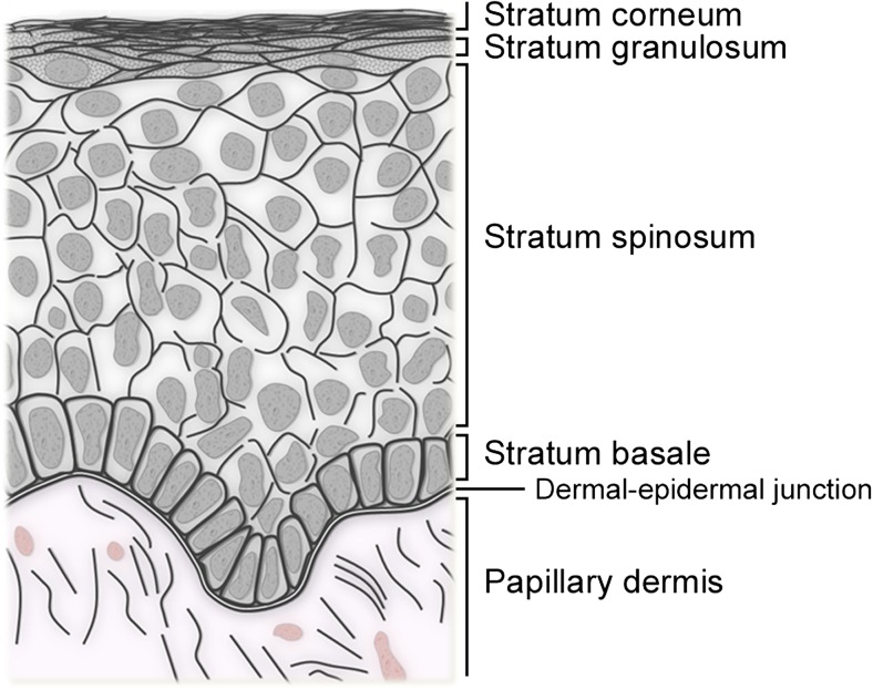
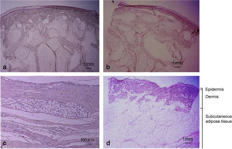
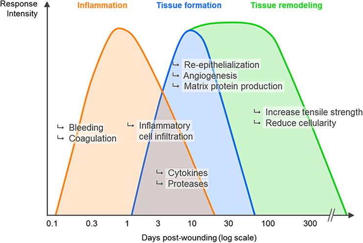
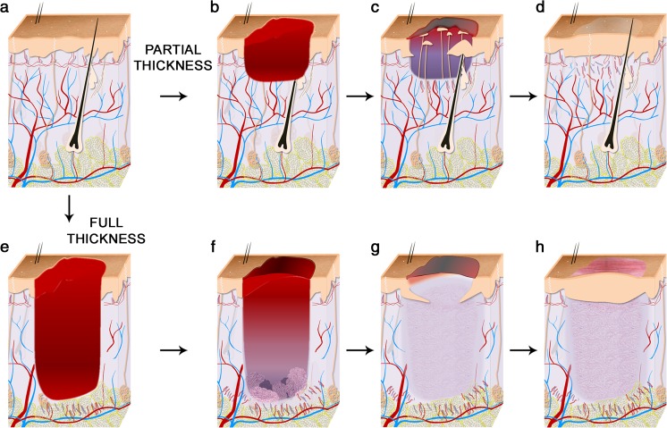
The increased incidence of non-healing skin wounds in developed societies has prompted tremendous research efforts on the complex process known as "wound healing". Unfortunately, the weak relevance of modern wound healing research to human health continues to be a matter of concern. This review summarizes the current knowledge of the cellular mechanisms that mediate wound closure in the skin of humans and laboratory animals. The author highlights the anatomical singularities of human skin vs. the skin of other mammals commonly used for wound healing research (i.e. as mice, rats, rabbits, and pigs), and discusses the roles of stem cells, myofibroblasts, and the matrix environment in the repair process. The majority of this review focuses on reepithelialization and wound closure. Other aspects of wound healing (e.g. inflammation, fibrous healing) are referred to when relevant to the main topic. This review aims at providing the reader with a clear understanding of the similarities and differences that have been reported over the past 100 years between the healing of human wounds and that of other mammals.

Keywords: Epidermis; Extracellular matrix; Granulation tissue; Mechanical force; Myofibroblast; Skin; Stem cells; Wound healing.


Figures

Fig. 1
Fig. 1
Organization of the human epidermis. The human skin epidermis is a stratified epithelium comprised of four levels (or strata) on most body sites. These strata are (from deep to superficial): (1) the basal layer (or stratum basale), a unique layer in unwounded skin that contains proliferative keratinocytes and inter-follicular keratinocyte stem cells; (2) the spinous layer (stratum spinosum) is made of several layers of polyhedral keratinocytes that have lost their proliferation potential and initiated differentiation; (3) the granular layer (stratum granulosum). Its granulated appearance in hematoxylin and eosin staining is due to the presence of kerato-hyalin granules, which are filled with proteins highly cross-linked with keratin filaments; and (4) the horny layer (stratum corneum), made of 15–20 layers of cornified (dead) cells devoid of nucleus or cytoplasmic organelles. In addition, a stratum lucidum can be distinguished between stratum granulosum and stratum corneum in the thick skin of the palms and soles. In older publications, the reader might commonly find the description of a Malpighian layer (which comprises stratum spinosum and stratum granulosum), prickle cells (cells of the stratum spinosum in which many desmosomes can be seen), or prickle cell layer (stratum spinosum)
Fig. 2
Fig. 2
Human subcutaneous adipose tissue varies in thickness and organization among skin sites. HE staining of excised skin obtained postmortem from the posterior aspect of heel (a), the plantar aspect of heel (b), the sacrum (c) and the center of the gluteus maximus (d). The three layers that comprise human skin are shown. Note that the adipose layer can be more than 1 cm-thick on some skin sites. Reproduced from (Arao et al. 2013) with permission
Fig. 3
Fig. 3
Phases of skin repair. The wound healing process is typically described as the succession of three overlapping phases: inflammation, tissue formation (or proliferation phase), and tissue remodeling. See text for details. This depiction derives mostly from the study of animal wounds and illustrates a wound repair process more segregated and less context-dependent than it is
Fig. 4
Fig. 4
Mechanisms of healing of partial- and full-thickness wounds in human skin. a Unwounded human skin is highly vascularized and contains hair follicles and eccrine sweat glands in a ratio 1:3 on most body sites. b–d Partial-thickness wounds involve the epidermis and may involve a portion of the dermis. They heal primarily by reepithelialization. New epidermis forms from outgrowths of eccrine sweat glands and pilosebaceous units underlying the wound. Keratinocyte migration from the wound edge is minimal. Wounds heal with minimal scarring and skin structure is minimally altered in the repaired site. e–g) Full-thickness wounds destroy the dermis (and possibly more). They do not heal by reepithelialization alone but require formation of a granulation tissue to fill the void of the wound before epithelial covering. After healing, the wound site bares a scar. Skin structure remains greatly modified with disappearance of appendages and altered vasculature
Fig. 5
Fig. 5
Organization of human granulation tissue as deducted from the work of Rosalind Butterworth (Butterworth 1992). a Schematic of the layered organization of the granulation tissue of large full-thickness human wounds healing by second intension. The ‘fibroblast’ layer is mostly populated by proto-myofibroblasts and myofibroblasts, and inflammatory cells are intravascular. Capillaries are present but they are few, large and mature. The capillary layer is the most cellular (see details in b). The loose connective tissue layer contains few, but viable, cells of varying type, and no endothelial cells or formed capillaries. The superficial slough contains fibrin and dead cells. Numbers to the right are average thickness of respective layer in μm. b Cellular composition of the capillary layer of human granulation tissue in the course of healing. Graphs were prepared according to raw data presented in (Butterworth 1992) from 80 biopsies. The cellularity of the capillary layer (the most cellular of all) remains fairly constant throughout healing

References

    1. Abe R, Donnelly SC, Peng T, Bucala R, Metz CN. Peripheral blood fibrocytes: differentiation pathway and migration to wound sites. J Immunol. 2001;166:7556–7562. doi: 10.4049/jimmunol.166.12.7556. - DOI - PubMed
    1. Arao H, Shimada T, Hagisawa S, Ferguson-Pell M. Morphological characteristics of the human skin over posterior aspect of heel in the context of pressure ulcer development. J Tissue Viability. 2013;22:42–51. doi: 10.1016/j.jtv.2013.02.002. - DOI - PubMed
    1. Arey LB. Certain basic principles of wound healing. Anat Rec. 1932;51:299–313. doi: 10.1002/ar.1090510309. - DOI
    1. Argenbright LW, Forbes PD. Erythema and skin blood content. Br J Dermatol. 1982;106:569–574. doi: 10.1111/j.1365-2133.1982.tb04560.x. - DOI - PubMed
    1. Arwert EN, Hoste E, Watt FM. Epithelial stem cells, wound healing and cancer. Nat Rev Cancer. 2012;12:170–180. doi: 10.1038/nrc3217. - DOI - PubMed