Review
. 2016 Jul;73(14):2809-19.
doi: 10.1007/s00018-016-2251-9. Epub 2016 May 12.

Starch-degrading polysaccharide monooxygenases

Affiliations
Review

Starch-degrading polysaccharide monooxygenases

Van V Vu et al. Cell Mol Life Sci. 2016 Jul.

Abstract

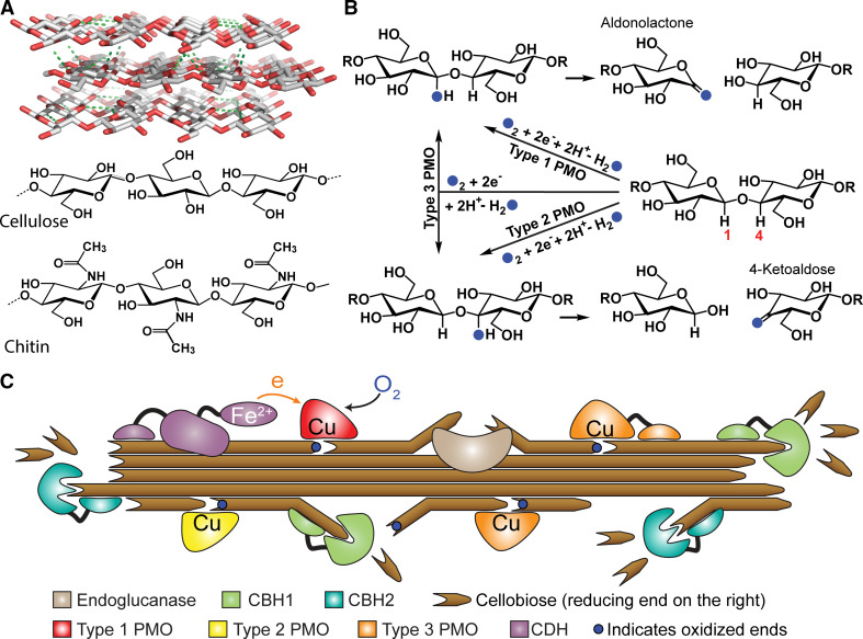
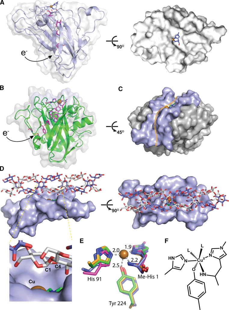
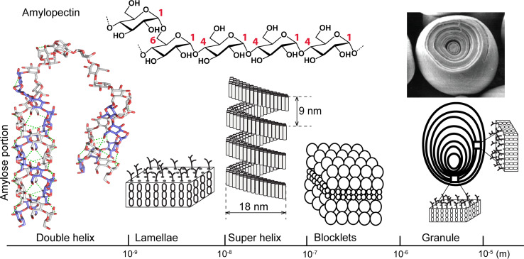
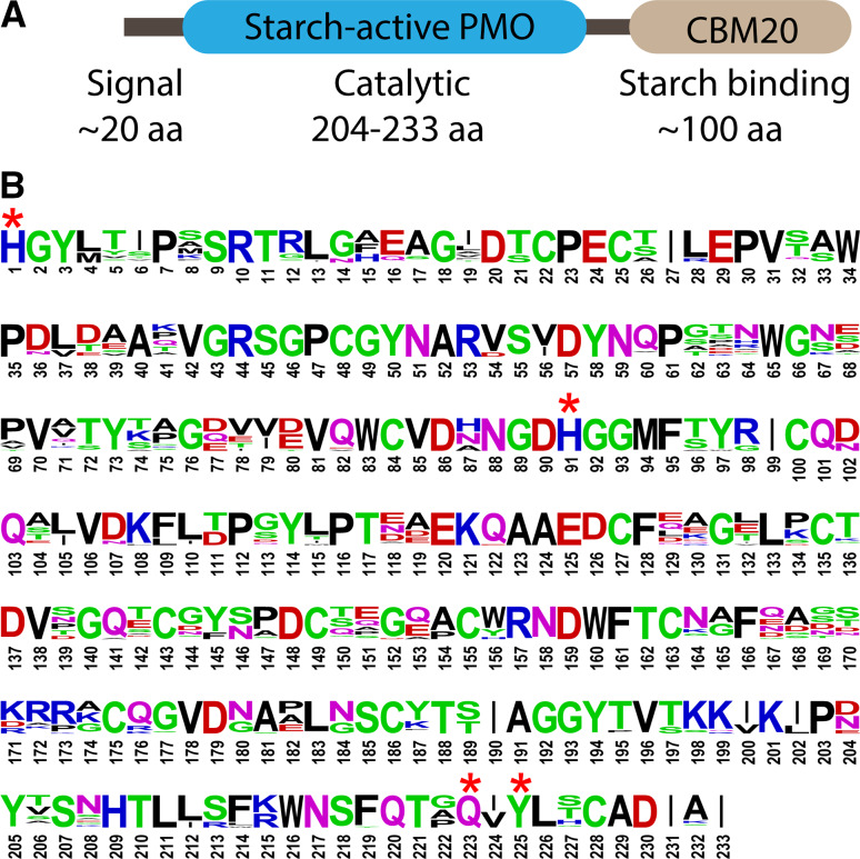
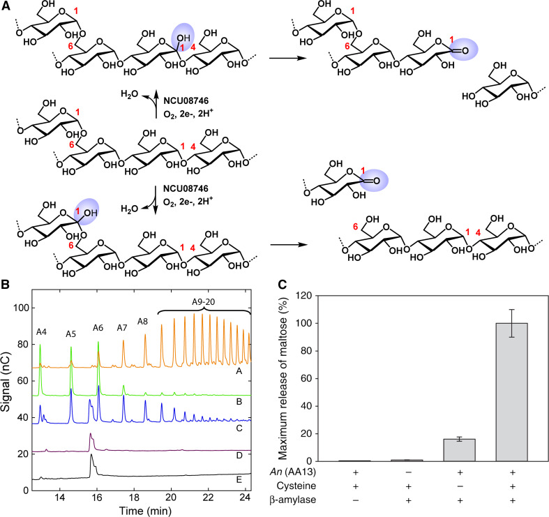
Polysaccharide degradation by hydrolytic enzymes glycoside hydrolases (GHs) is well known. More recently, polysaccharide monooxygenases (PMOs, also known as lytic PMOs or LPMOs) were found to oxidatively degrade various polysaccharides via a copper-dependent hydroxylation. PMOs were previously thought to be either GHs or carbohydrate binding modules (CBMs), and have been re-classified in carbohydrate active enzymes (CAZY) database as auxiliary activity (AA) families. These enzymes include cellulose-active fungal PMOs (AA9, formerly GH61), chitin- and cellulose-active bacterial PMOs (AA10, formerly CBM33), and chitin-active fungal PMOs (AA11). These PMOs significantly boost the activity of GHs under industrially relevant conditions, and thus have great potential in the biomass-based biofuel industry. PMOs that act on starch are the latest PMOs discovered (AA13), which has expanded our perspectives in PMOs studies and starch degradation. Starch-active PMOs have many common structural features and biochemical properties of the PMO superfamily, yet differ from other PMO families in several important aspects. These differences likely correlate, at least in part, to the differences in primary and higher order structures of starch and cellulose, and chitin. In this review we will discuss the discovery, structural features, biochemical and biophysical properties, and possible biological functions of starch-active PMOs, as well as their potential application in the biofuel, food, and other starch-based industries. Important questions regarding various aspects of starch-active PMOs and possible economical driving force for their future studies will also be highlighted.

Keywords: Auxiliary activity family 13; Biofuels; Copper enzymes; Plant pathogens; Polysaccharide monooxygenases; Starch degradation.


Figures

Fig. 1
Fig. 1
a Model of a cellulose crystal and the basic structure of cellulose and chitin. b Hydroxylation-mediated cleavage of the glycosidic bond by three types of cellulose-active PMOs. PMOs hydroxylate either C1 or C4 position of the glycosidic linkage, forming unstable intermediates that subsequently undergo elimination. Blue filled circles represent oxygen derived from O2. c Synergistic working model of PMOs with cellobiose dehydrogenase (CDH) and cellobiohydrolases (CBH1 and CBH2) in degrading cellulose (reproduced from Ref. [12] with permission)
Fig. 2
Fig. 2
Structure of starch active PMO in comparison with other PMO family and possible interaction with amylose substrate. a Structure of a cellulose-active PMO (4EIS) showing a flat active site surface. b Structure of starch-active PMO from A. oryzae, Ao(AA13), (4OPB) with similar β-sandwich core as found in other PMOs. Putative electron transfer pathways residues are displayed in magenta color in a and b. c Active site surface of Ao(AA13) exhibits a shallow groove. d Possible interaction of an amylose double helix with the active site groove of starch-active PMOs (manually docked). e XRD structure of Ao(AA13) (2OBP) active site (green) in comparison to that in an AA9 member (3ZUD, magenta, rmsd for protein atoms shown of 0.73 Å), AA10 member (2YOY, orange, rmsd of 0.53 Å) and AA11 member (4MAI, purple, rmsd of 0.60 Å) (reproduced from Ref. [11] with permission). f Solution structure deduced from XAS and UV/Vis spectroscopic analysis, where L can be aqueous or other coordinating agents in the solution
Fig. 3
Fig. 3
Primary structure of amylopectin, and higher order structures of starch on different size-scales. Amylose double helices form lamellae and superhelices. Blocklets contain one or more superhelices. Lamellae form crystalline growth rings that alternate with amorphous layers in starch granules. SEM image of starch and granule was reproduced from Ref. [31] with permission; Lamellae, super helix, blocklets, and granule were re-drawn based on Fig. 21 of Ref. [29] with permission
Fig. 4
Fig. 4
a Common domain architecture of 85 starch-active PMOs from different fungal species. Nineteen do not have the CBM20 domain [11]. b Consensus sequence logo representing the putative catalytic domain. Asterisks indicate the absolutely conserved residues also found in cellulose-active PMOs and chitin-active PMOs. This figure is reproduced from Ref. [10] with permission
Fig. 5
Fig. 5
a Proposed starch degradation steps by NCU08746 involving the cleavage of α(1 → 4) and α(1 → 6) linkages via hydroxylation at the C1 position. Further studies are required to determine whether starch-active PMO also oxidize α(1 → 6) linkage. b Activity assays of N. crassa starch-active PMOs, NCU08746, with ascorbic acid and atmospheric oxygen. Traces A and B maltoaldonic acids with four (A4) to twenty (A20) units. Traces C–E assays with amylopectin, phosphoric acid swollen cellulose, and chitin, respectively. c Boosting effect of AA13. Release of maltose from retrograded starch by β-amylase over 4 h at 25 °C (columns 2–4) with An(AA13) (column 3), with reducing agent and An(AA13) (column 4). a and b were reproduced from Ref. [10] and c from Ref. [11] with permission

References

    1. Vaaje-Kolstad G, Westereng B, Horn SJ, Liu Z, Zhai H, Sørlie M, Eijsink VG. An oxidative enzyme boosting the enzymatic conversion of recalcitrant polysaccharides. Science. 2010;330:219–222. doi: 10.1126/science.1192231. - DOI - PubMed
    1. Harris PV, Welner D, McFarland KC, Re E, Navarro Poulsen JC, Brown K, Salbo R, Ding H, Vlasenko E, Merino S, Xu F, Cherry J, Larsen S, Lo Leggio L. Stimulation of lignocellulosic biomass hydrolysis by proteins of glycoside hydrolase family 61: structure and function of a large, enigmatic family. Biochemistry. 2010;49:3305–3316. doi: 10.1021/bi100009p. - DOI - PubMed
    1. Quinlan RJ, Sweeney MD, Lo Leggio L, Otten H, Poulsen JC, Johansen KS, Krogh KB, Jørgensen CI, Tovborg M, Anthonsen A, Tryfona T, Walter CP, Dupree P, Xu F, Davies GJ, Walton PH. Insights into the oxidative degradation of cellulose by a copper metalloenzyme that exploits biomass components. Proc Natl Acad Sci USA. 2011;108:15079–15084. doi: 10.1073/pnas.1105776108. - DOI - PMC - PubMed
    1. Phillips CM, Beeson WT, Cate JHD, Marletta MA. Cellobiose dehydrogenase and a copper-dependent polysaccharide monooxygenase potentiate cellulose degradation by Neurospora crassa . ACS Chem Biol. 2011;6:1399–1406. doi: 10.1021/cb200351y. - DOI - PubMed
    1. Beeson WT, Phillips CM, Cate JHD, Marletta MA. Oxidative cleavage of cellulose by fungal copper-dependent polysaccharide monooxygenases. J Am Chem Soc. 2012;134:890–892. doi: 10.1021/ja210657t. - DOI - PubMed

Publication types