Review
. 2016 Nov;7(6):824-837.
doi: 10.1002/wrna.1361. Epub 2016 May 12.

Virus-derived small RNAs: molecular footprints of host-pathogen interactions

Affiliations
Review

Virus-derived small RNAs: molecular footprints of host-pathogen interactions

Eric Roberto Guimarães Rocha Aguiar et al. Wiley Interdiscip Rev RNA. 2016 Nov.

Abstract

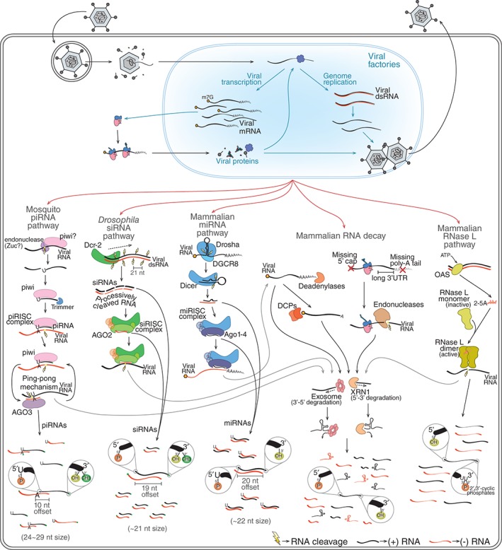
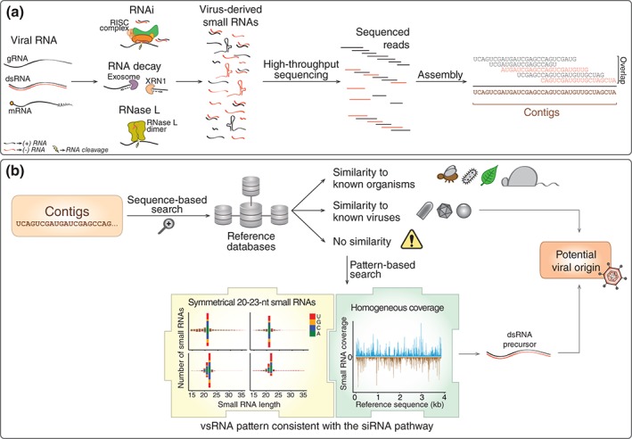
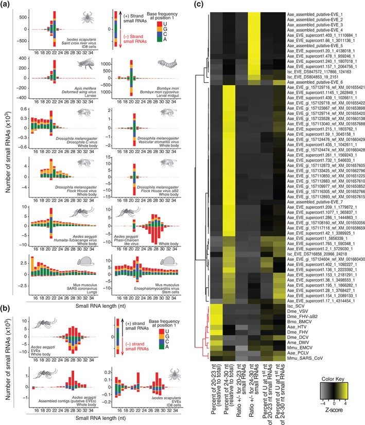
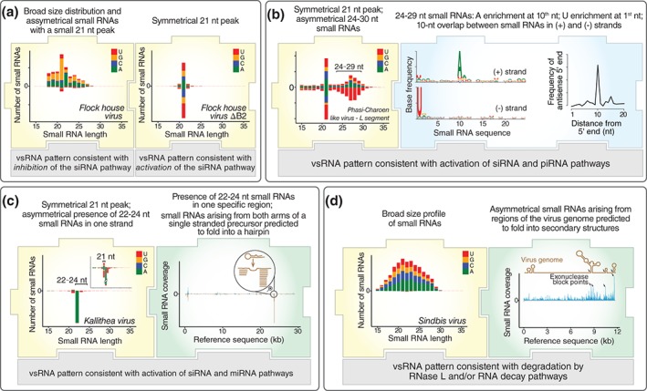
Viruses are obligatory intracellular parasites that require the host machinery to replicate. During their replication cycle, viral RNA intermediates can be recognized and degraded by different antiviral mechanisms that include RNA decay, RNA interference, and RNase L pathways. As a consequence of viral RNA degradation, infected cells can accumulate virus-derived small RNAs at high levels compared to cellular molecules. These small RNAs are imprinted with molecular characteristics that reflect their origin. First, small RNAs can be used to reconstruct viral sequences and identify the virus from which they originated. Second, other molecular features of small RNAs such as size, polarity, and base preferences depend on the type of viral substrate and host mechanism of degradation. Thus, the pattern of small RNAs generated in infected cells can be used as a molecular footprint to identify and characterize viruses independent on sequence homology searches against known references. Hence, sequencing of small RNAs obtained from infected cells enables virus discovery and characterization using both sequence-dependent strategies and novel pattern-based approaches. Recent studies are helping unlock the full application of small RNA sequencing for virus discovery and characterization. WIREs RNA 2016, 7:824-837. doi: 10.1002/wrna.1361 For further resources related to this article, please visit the WIREs website.


Figures

Figure 1
Figure 1
The generation of virus‐derived small RNAs by host pathways. During infection, virus genomes and transcripts are sometimes exposed and can be recognized by different host RNA surveillance mechanisms. RNA decay, RNA interference (RNAi), and RNase L pathways are good examples of such mechanisms. Mammalian RNA decay involves different mechanisms such as deadenylation‐dependent and nonsense‐mediated decay (NMD). The former involves shortening of the poly(A) tail by deadenylases followed by removal of the 5′ cap by mRNA‐decapping enzymes (DCPs). NMD is initiated by recognition of aberrant mRNAs that are cleaved by endonucleases. In both cases, the initial cleavage allows the mRNA to be targeted 5′→3′ by XRN1 and 3′→5′ by the exosome complex. The mammalian RNase L pathway is triggered when viral dsRNA is recognized by OAS enzymes that catalyze the production of 2′‐5′ oligoadenylates (2‐5A). These molecules induce dimerization and activation of RNase L that cleaves single‐stranded RNAs mostly at U‐rich regions. Different RNAi mechanisms can generate vsRNAs during viral infection including the mammalian miRNA pathway, Drosophila small interfering RNA (siRNA) pathway, and mosquito piRNA pathway. The Drosophila siRNA pathway is activated by Dcr‐2‐mediated recognition of viral dsRNA that is processes progressively to generate phased duplex siRNAs. siRNA duplexes are loaded onto AGO2 to generate siRISC that will find and cleave complementary RNAs. The mammalian miRNA pathway is initiated by the recognition of structure regions within long transcripts in the nucleus by the RNase III Drosha. This enzyme, in association with a partner protein known as DGCR8, cleaves the primary transcript to excise a short hairpin (~65 nt) that is then exported to the cytoplasm. There, the hairpin is further processed by Dicer to generate miRNA duplexes of ~22 nt that will be loaded onto different mammalian Argonaute proteins (Ago1–4) to form miRISC. This complex targets complementary regions within the 3′ UTR of mRNAs leading to translation inhibition. The mosquito piRNA pathway is triggered by the recognition of single‐stranded RNA precursors in a manner dependent on a specialized group of Argonautes known as PIWI proteins usually associated with an endonuclease known as Zucchini (Zuc). This initial recognition triggers processing of the precursor into primary piRNAs that remain associated with PIWI proteins to form the piRISC. This complex carries out cleavage of complementary RNA and can also initiate the production of more piRNAs. These secondary piRNAs require an amplification loop, referred to as the ping‐pong mechanism, that involves another Argonaute protein known as AGO3. Different RNA surveillance mechanisms may work together to generate vsRNAs. For example, RNA fragments generated by RNAi and RNase L pathways can be further targeted by RNA decay mechanisms. RNA surveillance mechanisms generate vsRNAs that have unique molecular characteristics such as terminal modifications, size, strand bias, and nucleotide preferences shown in the figure.
Figure 2
Figure 2
Identification of viral sequences utilizing virus‐derived small RNAs. (a) Virus‐derived small RNAs are generated by different host pathways during infection. These small RNAs can be sequenced and assembled into long contiguous sequences (contigs) reflecting the substrate from which they originate. (b) Classically, the origin of contigs from large‐scale sequencing studies is inferred by sequence similarities by comparisons to known references in databases. Thus, contigs are identified when they show similarity to sequences from known viruses or other organisms. However, a large number of contigs in large‐scale sequencing studies do not show similarity to any known reference sequence. In these cases, the molecular pattern of small RNAs derived from any contig sequence can be used. The presence of a clear small RNA symmetrical size preference between 20 and 23 nt at similar abundance on both strands of the contig suggests they originated from a double‐stranded RNA (dsRNA) precursor that was processed by the small interfering RNA (siRNA) pathway. This can be indicative of the contig origin because this molecular signature is reasonably specific to viruses. Thus, a small RNA profile can be used to identify potential viral contigs without any prior information about the sequence itself.
Figure 3
Figure 3
Activation of the small interfering RNA (siRNA) pathway is a common and specific response to virus infection. (a) Virus‐derived small RNAs in different animals often show a profile consistent with the activation of the siRNA pathway. Virus‐derived siRNAs range from ~20 to 23 nt and are symmetrical in polarity and base preferences. (b) Endogenous viral elements (EVEs) represent remnants of virus sequences integrated into animal genomes and also generate small RNAs. EVE‐derived small RNAs are often ~24–30 nt, asymmetrical in polarity, and base preferences that is consistent with a piRNA signature. (c) Size, polarity, and nucleotide preferences were determined for small RNAs derived from EVEs and active viruses. These molecular footprints were then used to perform hierarchical clustering of different EVEs and viruses. The small RNA pattern clearly separates clusters containing viruses (in red) and EVEs (in black). Notably, within the cluster of viruses, we observe two small subclusters. Virus grouped in the larger subcluster show a classical siRNA signature even when the pathway is partially inhibited such as for FHV and DCV in Drosophila. The second subcluster contains the vsRNA profile of coronavirus in mouse lungs and PCLV in mosquitoes, both of which show a more divergent profile from the siRNA signature. The data in this figure were obtained from the analysis of small RNA libraries from published studies (accession numbers: SRR1803378 , SRR1803382 , ERR555100, ERR274423, ERR654010, SRR1803383 , GSM792688, GSM792692, SRR452408, and SRR640612).10, 12, 77, 78, 79, 80, 81, 82
Figure 4
Figure 4
Molecular footprints of virus‐derived small RNAs. In addition to utilizing a classical small interfering RNA (siRNA) signature to identify potential viral sequences, the analysis of molecular characteristics of small RNAs derived from viral contigs can also provide further information about virus biology. The profile of virus‐derived small RNAs sometimes diverges from the canonical siRNA signature but still have unique molecular features that tend to reflect intrinsic properties of the virus such as tissue tropism, presence of inhibitors (VSR), and secondary structures or modifications in viral RNA. (a) The partial or complete absence of an expected siRNA signature often indicates that the virus escapes the siRNA pathway. This is the case of FHV in Drosophila, whose profile is fairly divergent from the canonical siRNA profiles unless a mutant virus incapable of producing the B2 VSR is analyzed. (b) The presence of longer virus‐derived small RNAs of ~24–30 nt that show a 10‐nt overlap between opposite strands is a signature of piRNAs. As these are often enriched in reproductive organs, this signature may be used to infer virus infection of ovaries as observed for PCLV in Aedes mosquitoes. (c) Animal viruses can sometimes show hotspots of small RNA coverage originating exclusively from one strand. These tend to come from genomic regions that are predicted to fold onto hairpins where the 20–24 nt long small RNAs arise from the stems. This small RNA signature suggests that this region is responsible for the generation of virus‐derived miRNAs as observed for Kallithea virus in Drosophila. (d) In the absence of a clear signature of the siRNA pathway, virus‐derived small RNAs may show a broad size distribution with highly heterogeneous coverage of the viral genome. This pattern suggests extensive viral RNA degradation with accumulation of small RNAs in regions corresponding to secondary structures or chemical modifications as observed for Sindbis virus in human cells. The small RNA data shown in this figure were obtained from the analysis of small RNA libraries from published studies (accession numbers GSM792688, GSM792692, SRR1803378 , SRR1914952 , and GSM1185388 ).10, 11, 61, 77

References

    1. den Boon JA, Diaz A, Ahlquist P. Cytoplasmic viral replication complexes. Cell Host Microbe 2010, 8:77–85. - PMC - PubMed
    1. Novoa RR, Calderita G, Arranz R, Fontana J, Granzow H, Risco C. Virus factories: associations of cell organelles for viral replication and morphogenesis. Biol Cell 2005, 97:147–172. - PMC - PubMed
    1. Marques JT, Carthew RW. A call to arms: coevolution of animal viruses and host innate immune responses. Trends Genet 2007, 23:359–364. - PubMed
    1. Ding S‐W. RNA‐based antiviral immunity. Nat Rev Immunol 2010, 10:632–644. - PubMed
    1. Chakrabarti A, Jha BK, Silverman RH. New insights into the role of RNase L in innate immunity. J Interferon Cytokine Res 2011, 31:49–57. - PMC - PubMed

Publication types

LinkOut - more resources