. 2016 Jun 24;90(14):6557-6572.
doi: 10.1128/JVI.00570-16. Print 2016 Jul 15.

Both CD8+ and CD4+ T Cells Contribute to Corneal Clouding and Viral Clearance following Vaccinia Virus Infection in C57BL/6 Mice

Affiliations

Both CD8+ and CD4+ T Cells Contribute to Corneal Clouding and Viral Clearance following Vaccinia Virus Infection in C57BL/6 Mice

I V Larsen et al. J Virol. .

Abstract

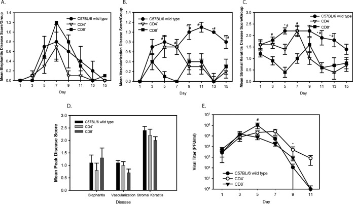
Vaccinia virus (VACV) keratitis is a serious complication following smallpox vaccination and can lead to blindness. The pathological mechanisms involved in ocular VACV infection are poorly understood. Previous studies have used rabbits, but the lack of immune reagents and transgenic or knockout animals makes them less suitable for mechanistic studies. We report that infection of C57BL/6 mice with 1 × 10(7) PFU of vaccinia virus strain WR results in blepharitis, corneal neovascularization, and stromal keratitis. The DryVax strain of VACV was completely attenuated. Infection required corneal scarification and replication-competent virus, and the severity of ocular disease was similar in 4- to 6-week-old and 1-year-old mice. Viral titers peaked at approximately 1 × 10(6) PFU on day 5 postinfection, and virus had not cleared by day 13 postinfection. Neutrophils were found in the peripheral cornea on day 1 after infection and then declined, followed by infiltration of both CD4(+) and CD8(+) T cells, which remained peripheral throughout the infection. Blood vessel growth extended 2 to 5 mm into the cornea from the limbus. Infection of CD4(-/-), CD8(-/-), or antibody-depleted mice resulted in similar disease severity and corneal clouding, indicating that both T-cell subsets were involved in the immunopathological response. Depletion of both CD4(+) and CD8(+) T cells resulted in significantly more severe disease and failure to clear the virus. On the basis of our results, the pathology of VACV keratitis is significantly different from that of herpes simplex virus keratitis. Further studies are likely to reveal novel information regarding virulence and immune responses to viral ocular infection.

Importance: Potentially blinding eye infections can occur after vaccination for smallpox. Very little is known about the pathological mechanisms that are involved, and the information that is available was generated using rabbit models. The lack of immunological reagents for rabbits makes such studies difficult. We characterized a mouse model of vaccinia virus ocular disease using C57BL/6 mice and strain WR and show that both CD4(+) and CD8(+) T-cell subsets play a role in the blinding eye disease and in controlling virus replication. On the basis of these results, vaccinia virus keratitis is significantly different from herpes simplex virus keratitis, and further studies using this model should generate novel insights into immunopathological responses to viral ocular infection.


Figures

FIG 1
FIG 1
Disease scores of vaccinia virus strain WR-infected C57BL/6 mice. (A) Blepharitis, vascularization, and stromal keratitis scores. The scores are the mean ± SEM per group. (B) Mean peak disease scores for blepharitis, vascularization, and stromal keratitis. Scores are the means of the highest scores for each mouse in a group ± SEMs. (C) Virus titers in tear film measured by plaque assay for C57BL/6 mice infected with the vaccinia virus WR strain. Titers are the mean number of PFU of virus per milliliter per group ± SEM.
FIG 2
FIG 2
Disease scores and viral titers for vaccinia virus strain WR-infected C57BL/6 mice at 6 weeks and 12 months of age. (A) Stromal keratitis scores, reported as the mean ± SEM per group. There were no significant differences between groups. (B) Mean peak disease scores for blepharitis, vascularization, and stromal keratitis. Scores are reported as the means of the highest scores for each mouse in a group ± SEMs. There were no significant differences between groups. (C) Virus titers in tear film measured by plaque assay for 6-week-old and 12-month-old infected mice. Titers are reported as the mean number of PFU of virus per milliliter for each group ± SEM. *, P < 0.05, day 1.
FIG 3
FIG 3
Disease scores and viral titers for C57BL/6 mice infected with vaccinia virus strain WR on unscarified corneas or infected using heat-inactivated virus and scarification. (A) Stromal keratitis scores, reported as the mean ± SEM per group. (B) Virus titers in tear films measured by plaque assay. Titers are reported as the mean number of PFU of virus per milliliter per group ± SEM. For days 1 through 9, the infected control mice had significantly higher stromal keratitis scores and titers than mice infected with inactivated virus or mice whose unscarified corneas were infected (P < 0.05).
FIG 4
FIG 4
(A to D) H&E staining of central and peripheral cornea. (A) Uninfected peripheral cornea, day 11; (B) infected peripheral cornea, day 7; (C) uninfected central cornea, day 11; (D) infected central cornea, day 11. (E to G) Immunostaining of infected peripheral cornea. Cells stained brown or red are positive. Nuclei counterstained with methyl green are negative. (E) Neutrophils in the peripheral cornea, day 1; (F) CD4+ cells in peripheral cornea, day 7; (G) CD8+ cells in peripheral cornea, day 9. (H) Staining with secondary antibody only. EP, corneal epithelium; S, stroma; EN, corneal endothelium.
FIG 5
FIG 5
Kinetics of cellular infiltration into infected corneas. The mean number of cells staining positive with antibodies specific for PMNs, CD8+ cells, and CD4+ cells in vaccinia virus-infected C57BL/6 mice is shown. Eyes were taken from three mice per group and sectioned, the number of positive cells in each section was counted, and the average number per group was determined.
FIG 6
FIG 6
Immunostaining of infected peripheral cornea for VACV antigen. Cells stained brown or red are positive for VACV. Nuclei were counterstained with hematoxylin. (A, B) Infected central and peripheral cornea, no primary antibody; (C, D) uninfected central and peripheral cornea; (E, F) infected central and peripheral cornea, eye with ocular disease, day 7. EP, corneal epithelium; S, stroma; EN, corneal endothelium; arrows, cells staining positive for viral antigen.
FIG 7
FIG 7
(A to C) Disease scores for vaccinia virus strain WR-infected C57BL/6 wild-type, CD4−/−, and CD8−/− mice. (A to C) Mean ± SEM blepharitis (A), vascularization (B), and stromal keratitis (C) scores per group. *, P < 0.05 for CD4−/− versus wild-type mice; #, P < 0.05 for CD8−/− versus wild-type mice. (D) Mean peak disease scores for blepharitis, vascularization, and stromal keratitis. Scores are the means of the highest scores for each mouse in a group ± SEMs. There were no significant differences between groups. (E) Virus titers in tear films measured by plaque assay for C57BL/6 wild-type, CD4−/−, and CD8−/− mice infected with the vaccinia virus WR strain. Titers are reported as the mean number of PFU of virus per milliliter per group ± SEM. *, P < 0.05 for CD4−/− versus wild-type mice, day 9; #, P < 0.05 for CD8−/− versus wild-type mice, day 5.
FIG 8
FIG 8
Histological analysis of central and peripheral corneas in wild-type, CD4−/−, and CD8−/− mice. Representative sections are shown. Sections were stained with hematoxylin and eosin. The dark pigment in the iris and ciliary body is melanin. The text above each panel indicates the group of mice represented and whether the image is from the central cornea or the periphery. EN, corneal endothelium; S, stroma; EP, corneal epithelium; CB, ciliary body; I, iris.
FIG 9
FIG 9
Flow cytometry analysis of T-cell subsets in infected mice in which CD4+ and CD8+ cells were depleted and not depleted. (A) Percentage of CD4+ and CD8+ cells in vaccinia virus WR-infected mice in which CD4+ cells, CD8+ cells, or both CD4+ and CD8+ cells were depleted, isotype control mice, and control C57BL/6 wild-type mice. Values are means ± SEMs. (B to H) Gating graphs for mice in which CD4+, CD8+, or CD4+ and CD8+ cells were depleted and isotype control and wild-type control mice in which CD4+ and CD8+ cells were not depleted. Pre Post, pre- and postinfection; Post, postinfection.
FIG 10
FIG 10
Disease scores of vaccinia virus strain WR-infected mice in which CD4+ cells were depleted (CD4 pre/post inf), CD8+ cells were depleted (CD8 pre/post inf), or both CD4+ and CD8+ cells were depleted (CD4/CD8 pre/post inf), in which CD4 cells were depleted postinfection (CD4 post inf), or in which CD8 cells were depleted postinfection (CD8 post inf), isotype control mice, and C57BL/6 mice in which CD4+ and CD8+ cells were not depleted (control). (A to C) Blepharitis (A), vascularization (B), and stromal keratitis (C) scores (mean ± SEM per group). *, P < 0.05 versus control group. (D) Mean peak disease scores for blepharitis, vascularization, and stromal keratitis. Scores are the means of the highest scores for each mouse in a group ± SEMs. *, P < 0.05 versus the control group. (E) Virus titers in tear films measured by plaque assay for vaccinia virus WR-infected mice in which CD4+ cells, CD8+ cells, or both CD4+ and CD8+ cells were depleted, isotype control mice, and C57BL/6 mice in which CD4+ and CD8+ cells were not depleted. Titers are reported as the mean number of PFU of virus per milliliter per group ± SEM. *, P < 0.05 versus the control group.
FIG 11
FIG 11
Histological analysis of central and peripheral corneas of antibody-depleted mice. Representative sections are shown. Sections were stained with hematoxylin and eosin. The dark pigment in the iris and ciliary body is melanin. The text above each panel indicates the group of mice represented and whether the image is from the central cornea or the periphery. EN, corneal endothelium; S, stroma; EP, corneal epithelium; CB, ciliary body; I, iris; depl pre/post, depletion of the indicated cell type pre- and postinfection; depl post, depletion of the indicated cell type postinfection; isotype, isotype control group.
FIG 12
FIG 12
Immunohistochemical staining of corneas from mice depleted of CD4+ T cells. Positive staining is indicated by the reddish brown color. The dark pigment in the iris and ciliary body is melanin. The text above each image indicates the location and the group. Note that the reddish brown color in the corneal epithelium and endothelium is background staining even with extensive blocking and peroxide treatment. EN, corneal endothelium; S, stroma; EP, corneal epithelium; CB, ciliary body; I, iris; arrows, cells positive for CD4+ cells.
FIG 13
FIG 13
Immunohistochemical staining of corneas of mice depleted of CD8+ T cells. Positive staining is indicated by the reddish brown color. The dark pigment in the iris and ciliary body is melanin. The text above each image indicates the location and the group. Note that the reddish brown color in the corneal epithelium and endothelium is background staining even with extensive blocking and peroxide treatment. EN, corneal endothelium; S, stroma; EP, corneal epithelium; CB, ciliary body; I, iris; arrows, cells positive for CD8+.

References

    1. Fenner F, Henderson DA, Arita I, Jezek Z, Ladnyi ID. 1988. Smallpox and its eradication. World Health Organization, Geneva, Switzerland.
    1. Quenelle DC, Kern ER. 2010. Treatment of vaccinia and cowpox virus infections in mice with CMX001 and ST-246. Viruses 2:2681–2695. doi: 10.3390/v2122681. - DOI - PMC - PubMed
    1. Croft DR, Sotir MJ, Williams CJ, Kazmierczak JJ, Wegner MV, Rausch D, Graham MB, Foldy SL, Wolters M, Damon IK, Karem KL, Davis JO. 2007. Occupational risks during a monkeypox outbreak, Wisconsin, 2003. Emerg Infect Dis 13:1150–1157. doi: 10.3201/eid1308.061365. - DOI - PMC - PubMed
    1. Lane JM, Ruben FL, Neff JM. 1969. Complications of smallpox vaccination. N Engl J Med 281:1201–1208. doi: 10.1056/NEJM196911272812201. - DOI - PubMed
    1. Ruben FL, Lane JM. 1970. Ocular vaccinia. An epidemiologic analysis of 348 cases. Arch Ophthalmol 84:45–48. - PubMed

Publication types