. 2016 Jul 11;90(15):6686-6698.
doi: 10.1128/JVI.00299-16. Print 2016 Aug 1.

Bile Acids Act as Soluble Host Restriction Factors Limiting Cytomegalovirus Replication in Hepatocytes

Affiliations

Bile Acids Act as Soluble Host Restriction Factors Limiting Cytomegalovirus Replication in Hepatocytes

Anna-Kathrin Schupp et al. J Virol. .

Abstract

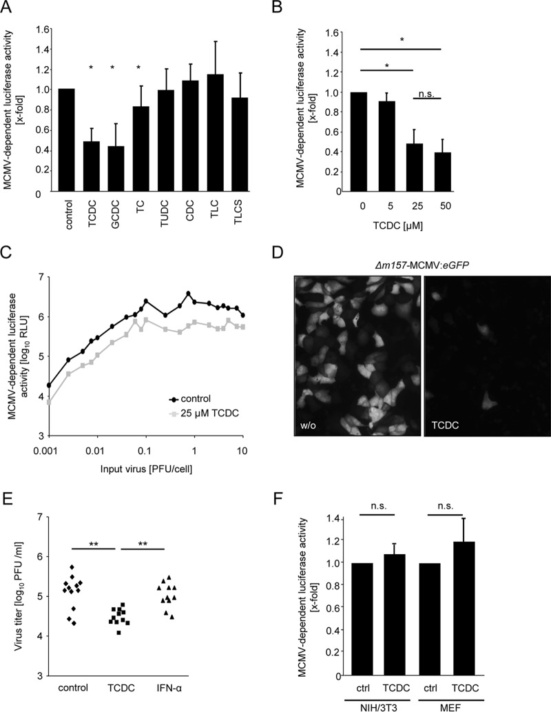
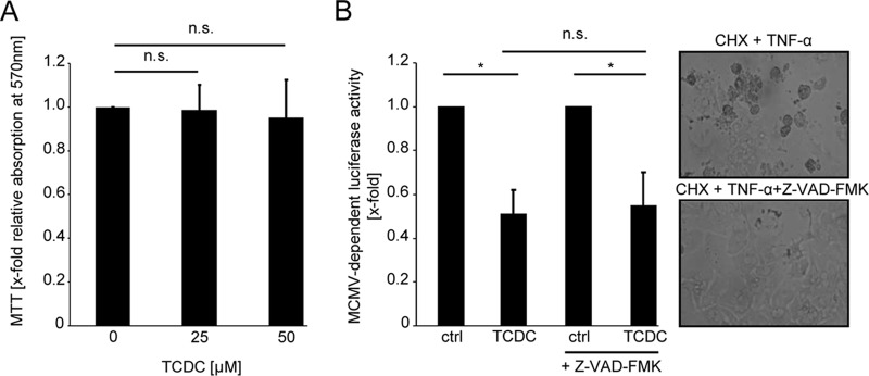
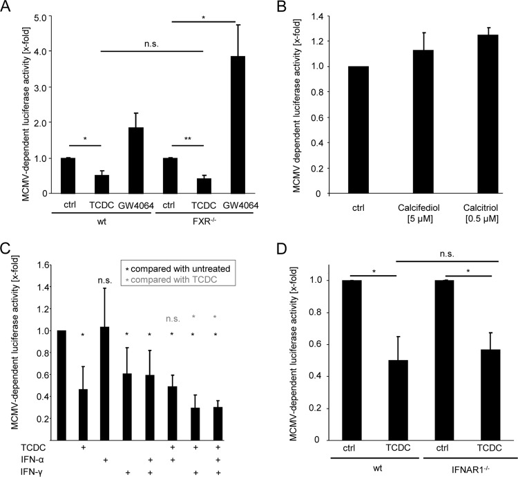
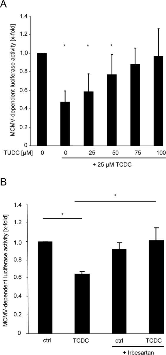
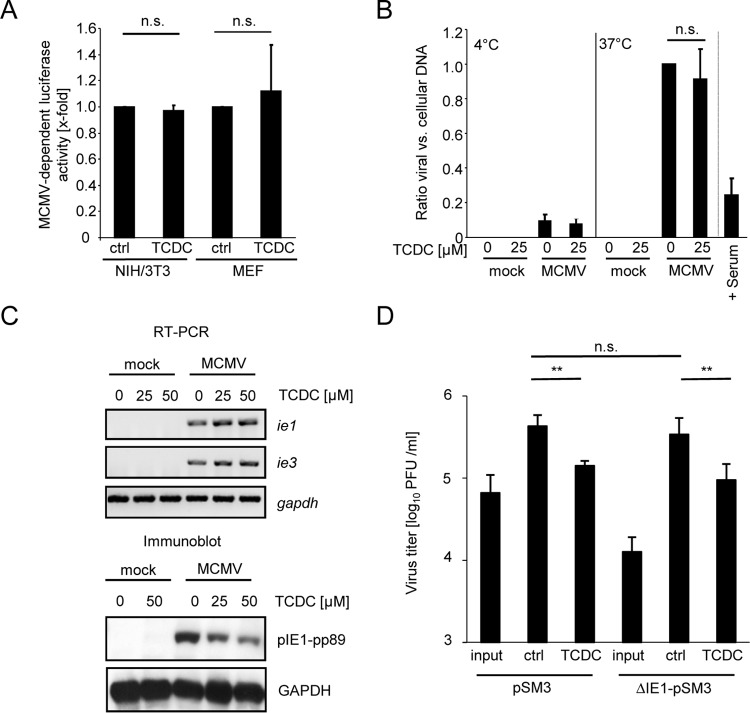
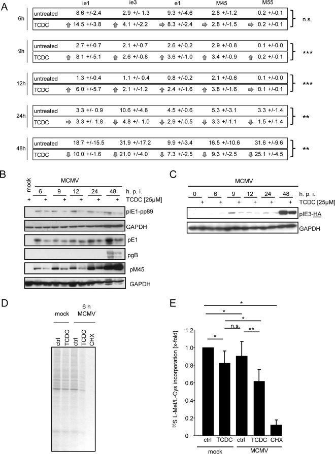
The liver constitutes a prime site of cytomegalovirus (CMV) replication and latency. Hepatocytes produce, secrete, and recycle a chemically diverse set of bile acids, with the result that interactions between bile acids and cytomegalovirus inevitably occur. Here we determined the impact of naturally occurring bile acids on mouse CMV (MCMV) replication. In primary mouse hepatocytes, physiological concentrations of taurochenodeoxycholic acid (TCDC), glycochenodeoxycholic acid, and to a lesser extent taurocholic acid significantly reduced MCMV-induced gene expression and diminished the generation of virus progeny, while several other bile acids did not exert antiviral effects. The anticytomegalovirus activity required active import of bile acids via the sodium-taurocholate-cotransporting polypeptide (NTCP) and was consistently observed in hepatocytes but not in fibroblasts. Under conditions in which alpha interferon (IFN-α) lacks antiviral activity, physiological TCDC concentrations were similarly effective as IFN-γ. A detailed investigation of distinct steps of the viral life cycle revealed that TCDC deregulates viral transcription and diminishes global translation in infected cells.

Importance: Cytomegaloviruses are members of the Betaherpesvirinae subfamily. Primary infection leads to latency, from which cytomegaloviruses can reactivate under immunocompromised conditions and cause severe disease manifestations, including hepatitis. The present study describes an unanticipated antiviral activity of conjugated bile acids on MCMV replication in hepatocytes. Bile acids negatively influence viral transcription and exhibit a global effect on translation. Our data identify bile acids as site-specific soluble host restriction factors against MCMV, which may allow rational design of anticytomegalovirus drugs using bile acids as lead compounds.


Figures

FIG 1
FIG 1
Conjugated bile acids act antivirally against mouse cytomegalovirus (MCMV). (A) Primary mouse hepatocytes were treated for 3 h with 25 μM concentrations of the bile acids taurochenodeoxycholic acid (TCDC), glycochenodeoxycholic acid (GCDC), taurocholic acid (TC), tauroursodeoxycholic acid (TUDC), chenodeoxycholic acid (CDC), taurolithocholic acid (TLC), and taurolithocholic sulfate (TLCS) and were infected with Δm157-MCMV-luciferase (0.1 PFU/cell) for the next 24 h thereafter. Hepatocytes were lysed, and luciferase activity was measured as described in Materials and Methods. The luciferase activity is calculated in relation to untreated control samples. (B) Primary mouse hepatocytes were incubated with the indicated TCDC concentrations for 3 h and subsequently infected with Δm157-MCMV-luciferase (0.1 PFU/cell) for the next 24 h. Cells were lysed, and the luciferase activity was determined. (C) Hepatocytes were incubated with 25 μM TCDC for 3 h and then infected with graded virus doses of Δm157-MCMV-luciferase (0.001 to 10 PFU/cell). Luciferase activity was measured at 24 h postinfection. (D) Hepatocytes were incubated with 25 μM TCDC for 3 h and infected with Δm157-MCMV-HMIEP-eGFP (0.1 PFU/cell) for the next 48 h. MCMV-driven eGFP expression was visualized by fluorescence microscopy. Representative fields are shown. (E) Primary mouse hepatocytes were treated with 25 μM TCDC or 500 U/ml IFN-α for 3 h and infected with Δm157-MCMV-HMIEP-eGFP (0.1 PFU/cell), and virus titers (in PFU/ml) were determined at 48 h postinfection by classical plaque titration. (F) NIH 3T3 and mouse embryonic fibroblast (MEF) cells were incubated for 3 h with 25 μM TCDC and subsequently infected with Δm157-MCMV-luciferase (0.1 PFU/cell). Luciferase activity was determined at 24 h postinfection.
FIG 2
FIG 2
TCDC does not inhibit CMV replication by inducing cytotoxicity. (A) Primary mouse hepatocytes were incubated for 48 h with 25 μM and 50 μM TCDC. A potential impact on cell viability was assessed by performing a 3-(4,5-dimethylthiazol-2-yl)-2,5-diphenyl-tetrazoliumbromide (MTT) assay. (B) To test the contribution of caspases to the antiviral activity of TCDC, primary mouse hepatocytes were conditioned with a 50 μM concentration of the pan-caspase inhibitor Z-VAD-FMK starting 30 min prior to the 3-h TCDC treatment (25 μM). The cells were infected with Δm157-MCMV-luciferase (0.1 PFU/cell) and lysed 24 h later for the determination of the luciferase activity. Functionality of the pan-caspase inhibitor was confirmed in parallel by coadministration with 100 μM cycloheximide (CHX) and 30 ng/ml TNF-α. Under this condition, Z-VAD-FMK effectively prevented caspase-dependent apoptosis (right panels).
FIG 3
FIG 3
Bile acids elicit their antiviral activity independently of FXR, vitamin D receptor, and IFNAR1. (A) Primary hepatocytes of FXR knockout and wild-type control mice were incubated with 25 μM TCDC or GW4064 for 3 h and subsequently infected with Δm157-MCMV-luciferase (0.1 PFU/cell) for the next 24 h before luciferase activity was measured. (B) Primary mouse hepatocytes were incubated for 24 h with 5 μM calcifediol or 0,5 μM calcitriol before being infected with Δm157-MCMV:luciferase (PFU:0.1). Luciferase activity was quantitated at 24 h after infection. (C) Primary mouse hepatocytes were conditioned for 24 h with 500 U/ml IFN-α, 500 U/ml IFN-γ, or a combination of both and, if indicated, for a further 3 h with 25 μM TCDC before the cells were infected with Δm157-MCMV-luciferase (0.1 PFU/cell) for an additional 24 h. Cells were lysed, and luciferase activity was quantified. (D) Primary hepatocytes of IFNAR1-deficient and wild-type mice were incubated for 3 h with 25 μM TCDC before infection with Δm157-MCMV-luciferase (0.1 PFU/cell). At 24 h postinfection, the cells were lysed and the luciferase activity was quantified.
FIG 4
FIG 4
Inhibition of MCMV replication by TCDC is dependent on NTCP-mediated cellular uptake. (A) Primary murine hepatocytes were conditioned for 3 h with 25 μM TCDC. Simultaneously, the indicated concentrations (0 to 100 μM) of TUDC were also added to the cells prior to infection with Δm157-MCMV-luciferase. Note that TUDC does not possess antiviral activity against MCMV (Fig. 1A). At 24 h postinfection, the cells were lysed and luciferase activity was quantified. (B) Primary mouse hepatocytes were incubated for 3 h with 25 μM TCDC, and 50 μM irbesartan was coadministered prior to infection with Δm157-MCMV-luciferase (0.1 PFU/cell). Luciferase activity was quantified at 24 h postinfection.
FIG 5
FIG 5
TCDC reduces IE1 protein amounts, but the antiviral effect of TCDC is independent of IE1 function. (A) Purified MCMV particles were incubated with 25 μM TCDC for 3 h at 37°C and were used for infection of NIH 3T3 and MEF cells. Note that both fibroblast cell types do not support the antiviral activity elicited by bile acids in/on hepatocytes (Fig. 1F). At 24 h postinfection, luciferase activity was quantified. (B) Copy numbers of intracellular genomic viral DNA in relation to cellular DNA were determined by quantitative TaqMan PCR with primers and probes specific for the viral gene region M92 and the cellular β-actin gene. Primary mouse hepatocytes were incubated for 3 h with 25 μM TCDC and then infected with MCMV (0.1 PFU/cell) either at 4°C (which allows virus attachment but precludes virus entry) or at 37°C (which allows virus entry). At 2 h postinfection, extracellular virus particles were removed by multiple rounds of vigorous trypsin washing. Subsequently, the cells were collected and the total (intra)cellular DNA was purified. An incubation of virus with immune serum of MCMV-infected mice (which is known to contain effective concentrations of neutralizing antibodies) served as a positive control for an inhibition of virus entry. (C) Primary mouse hepatocytes were incubated for 3 h with 25 μM and 50 μM TCDC and subsequently infected with MCMV for a further 6 h. Cells were lysed, and RNA was isolated. The abundance of immediate early (IE) ie1 and ie3 mRNAs and housekeeping GAPDH mRNA was determined by semiquantitative one-step reverse transcription-PCR with specific primers. In parallel, cells were lysed and protein lysates were prepared, normalized according to Bradford protein staining, separated by SDS-polyacrylamide gel electrophoresis, and subjected to immunoblotting followed by detection with the indicated antibodies. (D) Primary hepatocytes were treated for 3 h with 25 μM TCDC and infected with MCMV strain pSMA3 or ΔIE1-pSMA3 (0.1 PFU/cell) for 48 h. Virus titers (in PFU/ml) were determined by classic plaque titration.
FIG 6
FIG 6
TCDC inhibits MCMV replication at the level of protein translation and modifies viral mRNA expression. (A) Primary mouse hepatocytes were incubated with 25 μM TCDC for 3 h and infected with Δm157-MCMV-luciferase afterwards. Cells were lysed after 6 h, 9 h, 12 h, 24 h, or 48 h, and RNA was isolated. cDNA was synthesized, and quantitative PCR was performed with specific primers for ie1, ie3, early1, M45, and M55. Depicted are the relative RNA expression levels in comparison to that of the cellular transcript sdha. To calculate statistical significance, the results of all transcription values at one time point were considered (except in case of M55, where no gene expression was detectable in the 6- to 12-h period and only later time points were included in the analysis). (B) Primary hepatocytes were treated for 3 h with 25 μM TCDC and infected with MCMV-IE3-HA or Δm157-MCMV-luciferase (multiplicity of infection of 0.1). Hepatocytes were lysed at the indicated time points and pIE1, pIE3-HA, pE1, pM45, gB, and housekeeping GAPDH were detected by SDS-PAGE and immunoblotting with specific antibodies. (C) Primary mouse hepatocytes were preincubated for 3 h with 25 μM TCDC and infected with MCMV (1 PFU/cell). 35S-labeled l-Met/l-Cys (27 μCi) was added to the cells directly after centrifugally enhanced infection. After 6 h, cells were lysed with lysis buffer, proteins were separated by SDS-PAGE, the gel was fixed and dried, and the intensity of radioactively labeled proteins was visualized by autoradiography. The inhibitor of translation CHX was used as a positive control. (D) Intensities of 35S incorporation were quantified (for details, see Materials and Methods). The intensities were calculated in relation to untreated control samples.

References

    1. Rafailidis PI, Mourtzoukou EG, Varbobitis IC, Falagas ME. 2008. Severe cytomegalovirus infection in apparently immunocompetent patients: a systematic review. Virol J 5:47. doi:10.1186/1743-422X-5-47. - DOI - PMC - PubMed
    1. Ten Napel CH, Houthoff HJ, The TH. 1984. Cytomegalovirus hepatitis in normal and immune compromised hosts. Liver 4:184–194. - PubMed
    1. Mocarski ES, Shenk T, Pass RF. 2007. Cytomegaloviruses, p 2656–2701. In Knipe DM, Howley PM (ed), Fields virology, 5th ed, vol 2 Lippincott Williams & Wilkins, Philadelphia, PA.
    1. Buckner FS, Pomeroy C. 1993. Cytomegalovirus disease of the gastrointestinal tract in patients without AIDS. Clin Infect Dis 17:644–656. doi:10.1093/clinids/17.4.644. - DOI - PubMed
    1. Seehofer D, Rayes N, Tullius SG, Schmidt CA, Neumann UP, Radke C, Settmacher U, Muller AR, Steinmuller T, Neuhaus P. 2002. CMV hepatitis after liver transplantation: incidence, clinical course, and long-term follow-up. Liver Transpl 8:1138–1146. doi:10.1053/jlts.2002.36732. - DOI - PubMed

Publication types

MeSH terms