. 2016 Jul 11;90(15):6724-6737.
doi: 10.1128/JVI.00573-16. Print 2016 Aug 1.

Arteriviruses, Pegiviruses, and Lentiviruses Are Common among Wild African Monkeys

Affiliations

Arteriviruses, Pegiviruses, and Lentiviruses Are Common among Wild African Monkeys

Adam L Bailey et al. J Virol. .

Abstract

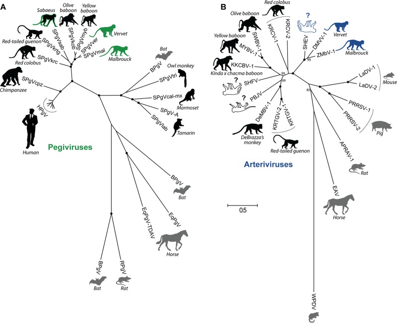
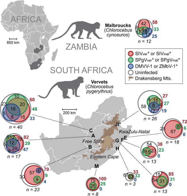
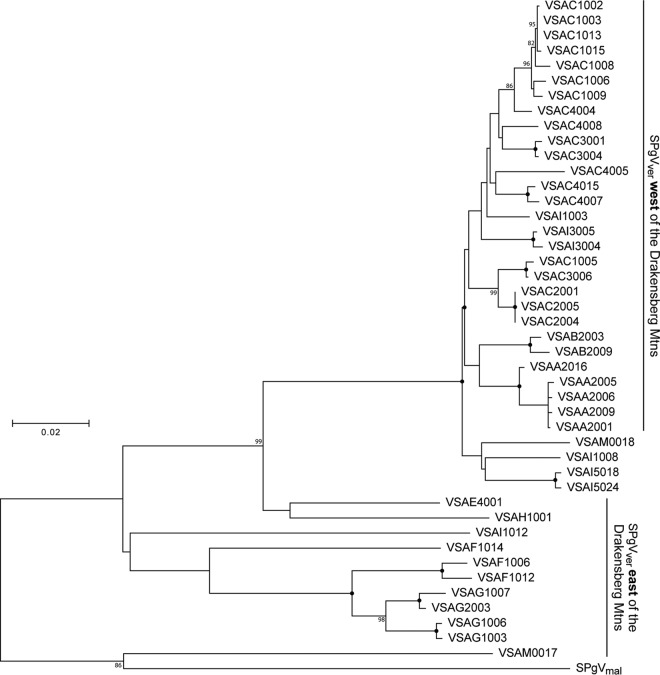
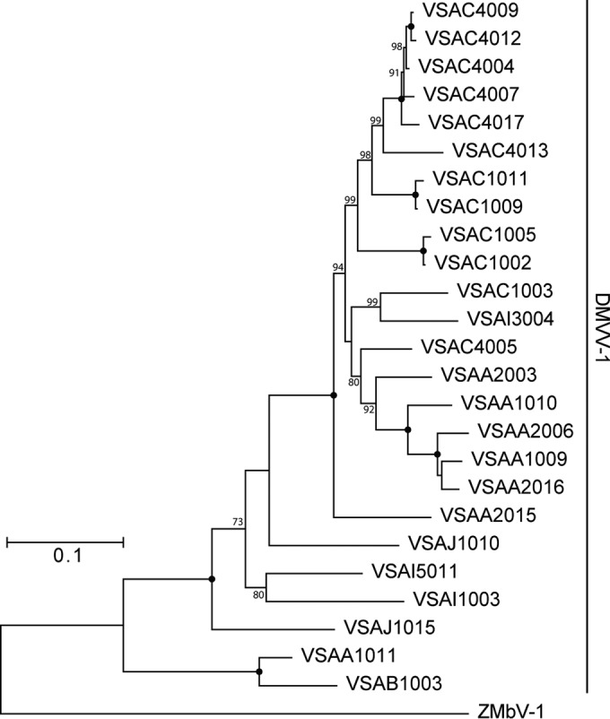
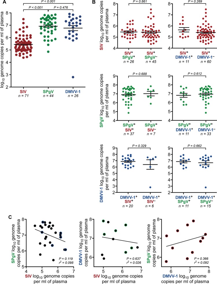
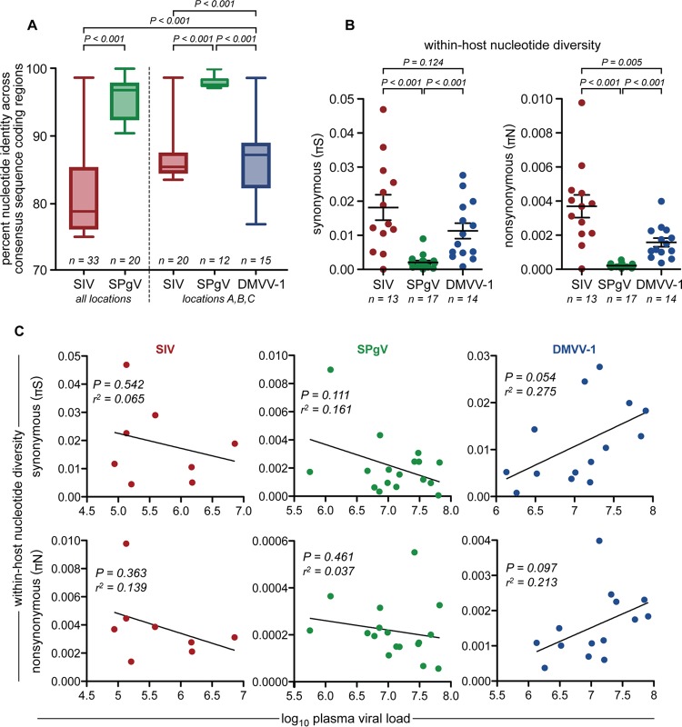
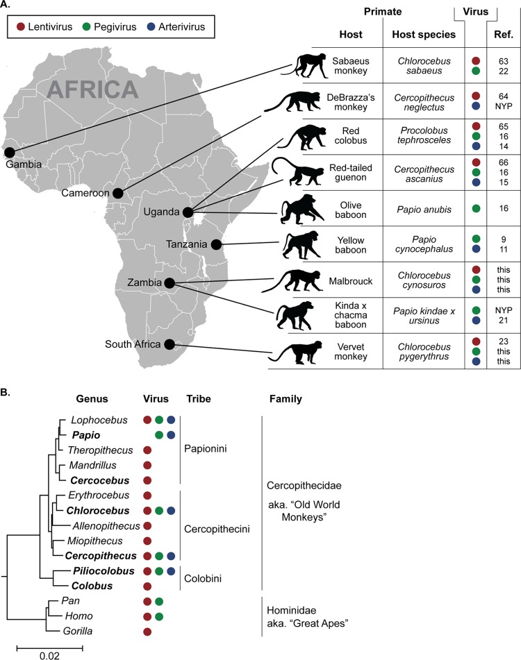
Nonhuman primates (NHPs) are a historically important source of zoonotic viruses and are a gold-standard model for research on many human pathogens. However, with the exception of simian immunodeficiency virus (SIV) (family Retroviridae), the blood-borne viruses harbored by these animals in the wild remain incompletely characterized. Here, we report the discovery and characterization of two novel simian pegiviruses (family Flaviviridae) and two novel simian arteriviruses (family Arteriviridae) in wild African green monkeys from Zambia (malbroucks [Chlorocebus cynosuros]) and South Africa (vervet monkeys [Chlorocebus pygerythrus]). We examine several aspects of infection, including viral load, genetic diversity, evolution, and geographic distribution, as well as host factors such as age, sex, and plasma cytokines. In combination with previous efforts to characterize blood-borne RNA viruses in wild primates across sub-Saharan Africa, these discoveries demonstrate that in addition to SIV, simian pegiviruses and simian arteriviruses are widespread and prevalent among many African cercopithecoid (i.e., Old World) monkeys.

Importance: Primates are an important source of viruses that infect humans and serve as an important laboratory model of human virus infection. Here, we discover two new viruses in African green monkeys from Zambia and South Africa. In combination with previous virus discovery efforts, this finding suggests that these virus types are widespread among African monkeys. Our analysis suggests that one of these virus types, the simian arteriviruses, may have the potential to jump between different primate species and cause disease. In contrast, the other virus type, the pegiviruses, are thought to reduce the disease caused by human immunodeficiency virus (HIV) in humans. However, we did not observe a similar protective effect in SIV-infected African monkeys coinfected with pegiviruses, possibly because SIV causes little to no disease in these hosts.


Figures

FIG 1
FIG 1
Species-level phylogenetic relationships of all known pegiviruses (A) and arteriviruses (B). Viruses are shown adjacent to the silhouette of their respective host, with host common names in italics. Primate hosts are shown in black, and nonprimate hosts are shown in gray. Viruses discovered in this study are depicted by host silhouettes with solid coloring: green for AGM pegiviruses and blue for AGM arteriviruses. Host silhouettes with a colored outline draw attention to viruses of importance. Sabaeus SPgV (SPgVsab) has a green outline because this virus does not group with the AGM pegiviruses presented here. SHEV has a blue outline because of its close relationship to the AGM arteriviruses presented here. White host silhouettes symbolize arteriviruses that have caused outbreaks of viral hemorrhagic fever in captive macaques. Question marks emphasize that the natural host(s) of these viruses remains unknown. Shown is a maximum likelihood tree with 1,000 bootstrap replicates. Black dots indicate splits that are supported by 100% of bootstrap replicates. Bootstrap values below 70 are not shown. The bar shows the calculated genetic distance. SPgVkrc, Kibale red colobus; SPgVkrtg, Kibale red-tailed guenon; SPgVob, SPgV from olive baboon; SPgVmyb, Mikumi yellow baboon; BPgV, bat pegivirus; SPgVtri, owl monkey; SPgVcal-mx, marmoset-mystax; SPgVlab, tamarin; EqPgV, equine pegivirus; TDAV, Theiler's disease-associated virus; RPgV, rat PgV; KRTGV-1, Kibale red-tailed guenon virus; DeMBV-1, Debrazza's monkey virus; PBJV, Peter B. Jahrling virus; KKCBV-1, Kafue kinda-chacma baboon virus; MYBV-1, Mikumi yellow baboon virus; SWBV-1, Southwest baboon virus; KRCV-1, Kibale red colobus virus; LaDV-1, lactate dehydrogenase elevating virus; PRRSV-1, porcine reproductive and respiratory syndrome virus; APRAV-1, African pouched rat virus; EAV, equine arteritis virus; WPDV, wobbly possum disease virus.
FIG 2
FIG 2
Geographic distribution and prevalence of AGM plasma viruses. Shown are prevalences of SIV, SPgV, and simian arteriviruses in wild AGMs sampled from Zambia (malbrouck monkey [Chlorocebus cynosuros]) and South Africa (vervet monkey [Chlorocebus pygerythrus]). Venn diagrams of each monkey population show the percentages of uninfected, monoinfected, coinfected, and triple-infected monkeys for each of these three viruses. Gray circles represent the total numbers of monkeys sampled and are proportional to the sample size from each location. Numbers within the gray circles but outside the colored circles are the percentages of each population that are triple negative for virus (white). Colored circles within gray circles show the percentages of each population infected with SIV, SPgV, or DMVV-1/ZMbV-1 that are monoinfected, coinfected, or triple infected. Adjacent colored numbers outside the circles show the percentages of each population infected with the respective virus. Sites I and J are from the Riverside Rehabilitation Center, which houses vervet monkeys from across the region. The prevalence of each virus was determined by using a combination of deep sequencing, qRT-PCR, and RT-PCR.
FIG 3
FIG 3
Phylogenetic relationships of all SPgV variants discovered in this study. A PCR amplicon spanning the putative NS3 coding region of the SPgVver genome was sequenced from samples that tested positive for SPgV RNA by qRT-PCR that were not subjected to unbiased deep sequencing. Shown is a maximum likelihood tree, not rooted, with 1,000 bootstrap replicates. Black dots indicate splits that are supported by 100% of bootstrap replicates. Bootstrap values below 70 are not shown. The bar shows the calculated genetic distance.
FIG 4
FIG 4
Phylogenetic relationships of all AGM simian arteriviruses discovered in this study. A PCR amplicon spanning ORF1b of the DMVV-1 genome was sequenced from samples that tested positive for DMVV-1 RNA by qRT-PCR that were not subjected to unbiased deep sequencing. Shown is a maximum likelihood tree, not rooted, with 1,000 bootstrap replicates. Black dots indicate splits that are supported by 100% of bootstrap replicates. Bootstrap values below 70 are not shown. The bar shows the calculated genetic distance.
FIG 5
FIG 5
Viremia of the AGM plasma viruses. (A) SPgVver and DMVV-1 RNA concentrations were quantified from plasma samples of 161 South African vervet monkeys by using highly sensitive virus-specific qRT-PCR assays designed from deep-sequencing data. Only positive results are shown. SIVver loads were determined previously (23) by the same method. Significance was assessed by using a two-tailed unpaired t test on log-transformed values, with error bars showing the standard errors of the means. (B) South African vervets were stratified by coinfection status, and plasma viral load values for the virus in question were plotted. Significance was assessed by using a two-tailed unpaired t test on log-transformed values, with error bars showing the standard errors of the means. (C) Linear regression correlating the viral load of each virus in coinfected individuals. Data points with a colored halo indicate triple-infected individuals.
FIG 6
FIG 6
Genetic diversity of AGM plasma viruses. (A) Complete consensus sequences spanning the entire coding region of each virus were aligned, and a pairwise comparison between each aligned sequence was performed. Percent identity values from each comparison were plotted and compared by using a two-tailed unpaired t test. Boxes show the middle two quartiles, and whiskers show the minimum and maximum percent identities observed. (B) Synonymous (πS) and nonsynonymous (πN) viral nucleotide diversities within each infected monkey, determined by calculating πS and πN values across the entire viral genome. Only samples that yielded virus sequences with >100× coverage for >99% of the protein-coding region of the genome were used for this analysis. Significance was assessed by using a two-tailed unpaired t test, with error bars showing the standard errors of the means. (C) Linear regression correlating within-host nucleotide diversity and plasma viral load for each virus.
FIG 7
FIG 7
Known geographic and host ranges of African monkey plasma viruses. (A) Map of Africa showing the sampling locations of monkeys in which simian pegivirus or simian arterivirus infection was identified. In the table, a colored dot indicates that SIV (red), SPgV (green), or a simian arterivirus (blue) infection was detected in that particular primate from that particular location (61–64). NYP, not yet published. (B) Genus-level phylogenetic tree of African OWMs and great apes. Colored dots indicate that SIV (red), GBV-C (green), or SHFV (blue) infection was detected in a primate from that genus. Names in boldface type indicate genera from which we have sampled more than 10 wild primates by unbiased deep sequencing (for a comprehensive list of species naturally infected with SIV, see reference 65). (Adapted from PLoS Genetics [66].)

References

    1. Wolfe ND, Dunavan CP, Diamond J. 2007. Origins of major human infectious diseases. Nature 447:279–283. doi: 10.1038/nature05775. - DOI - PMC - PubMed
    1. Davies TJ, Pedersen AB. 2008. Phylogeny and geography predict pathogen community similarity in wild primates and humans. Proc Biol Sci 275:1695–1701. doi: 10.1098/rspb.2008.0284. - DOI - PMC - PubMed
    1. Apetrei C, Robertson DL, Marx PA. 2004. The history of SIVS and AIDS: epidemiology, phylogeny and biology of isolates from naturally SIV infected non-human primates (NHP) in Africa. Front Biosci 9:225–254. doi: 10.2741/1154. - DOI - PubMed
    1. Sharp PM, Hahn BH. 2011. Origins of HIV and the AIDS pandemic. Cold Spring Harb Perspect Med 1:a006841. doi: 10.1101/cshperspect.a006841. - DOI - PMC - PubMed
    1. Chahroudi A, Bosinger SE, Vanderford TH, Paiardini M, Silvestri G. 2012. Natural SIV hosts: showing AIDS the door. Science 335:1188–1193. doi: 10.1126/science.1217550. - DOI - PMC - PubMed

Publication types

MeSH terms