

Noninvasive Real-Time Automated Skin Lesion Analysis System for Melanoma Early Detection and Prevention
- PMID: 27170906
- PMCID: PMC4848099
- DOI: 10.1109/JTEHM.2015.2419612
Noninvasive Real-Time Automated Skin Lesion Analysis System for Melanoma Early Detection and Prevention
Abstract
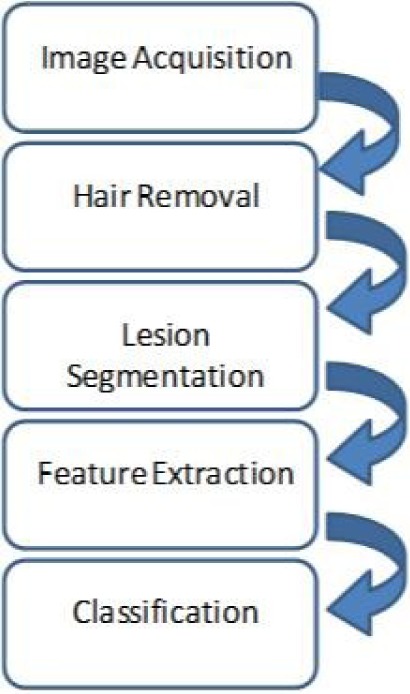
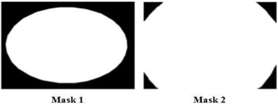
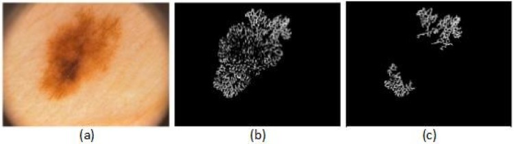
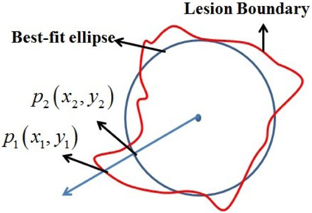
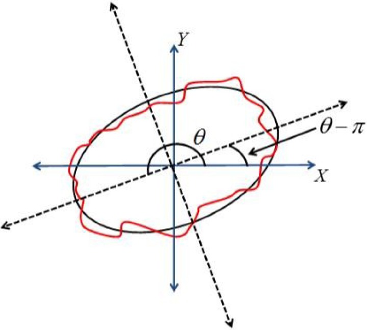
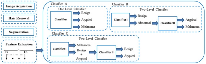
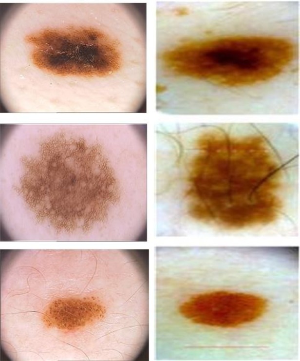
Melanoma spreads through metastasis, and therefore, it has been proved to be very fatal. Statistical evidence has revealed that the majority of deaths resulting from skin cancer are as a result of melanoma. Further investigations have shown that the survival rates in patients depend on the stage of the cancer; early detection and intervention of melanoma implicate higher chances of cure. Clinical diagnosis and prognosis of melanoma are challenging, since the processes are prone to misdiagnosis and inaccuracies due to doctors' subjectivity. Malignant melanomas are asymmetrical, have irregular borders, notched edges, and color variations, so analyzing the shape, color, and texture of the skin lesion is important for the early detection and prevention of melanoma. This paper proposes the two major components of a noninvasive real-time automated skin lesion analysis system for the early detection and prevention of melanoma. The first component is a real-time alert to help users prevent skinburn caused by sunlight; a novel equation to compute the time for skin to burn is thereby introduced. The second component is an automated image analysis module, which contains image acquisition, hair detection and exclusion, lesion segmentation, feature extraction, and classification. The proposed system uses PH2 Dermoscopy image database from Pedro Hispano Hospital for the development and testing purposes. The image database contains a total of 200 dermoscopy images of lesions, including benign, atypical, and melanoma cases. The experimental results show that the proposed system is efficient, achieving classification of the benign, atypical, and melanoma images with accuracy of 96.3%, 95.7%, and 97.5%, respectively.
Keywords: Image segmentation; melanoma; skin cancer.
Figures
References
-
- Rademaker M. and Oakley A., “Digital monitoring by whole body photography and sequential digital dermoscopy detects thinner melanomas,” J. Primary Health Care, vol. 2, no. 4, pp. 268–272, 2010. - PubMed
-
- Abuzaghleh O., Barkana B. D., and Faezipour M., “SKINcure: A real time image analysis system to aid in the malignant melanoma prevention and early detection,” in Proc. IEEE Southwest Symp. Image Anal. Interpretation (SSIAI), Apr. 2014, pp. 85–88.
-
- Abuzaghleh O., Barkana B. D., and Faezipour M., “Automated skin lesion analysis based on color and shape geometry feature set for melanoma early detection and prevention,” in Proc. IEEE Long Island Syst., Appl. Technol. Conf. (LISAT), May 2014, pp. 1–6.
-
- (Mar. 27, 2014). American Cancer Society, Cancer Facts & Figures. [Online]. Available: http://www.cancer.org/research/cancerfactsstatistics/cancerfactsfigures2...
LinkOut - more resources
Full Text Sources
Other Literature Sources
