. 2016 May 12;11(5):e0155420.
doi: 10.1371/journal.pone.0155420. eCollection 2016.

Interferon Regulator Factor 8 (IRF8) Limits Ocular Pathology during HSV-1 Infection by Restraining the Activation and Expansion of CD8+ T Cells

Affiliations

Interferon Regulator Factor 8 (IRF8) Limits Ocular Pathology during HSV-1 Infection by Restraining the Activation and Expansion of CD8+ T Cells

Lin Sun et al. PLoS One. .

Abstract

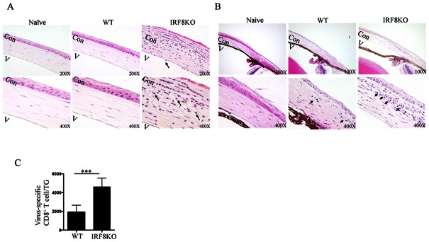
Interferon Regulatory Factor-8 (IRF8) is constitutively expressed in monocytes and B cell lineages and plays important roles in immunity to pathogens and cancer. Although IRF8 expression is induced in activated T cells, the functional relevance of IRF8 in T cell-mediated immunity is not well understood. In this study, we used mice with targeted deletion of Irf8 in T-cells (IRF8KO) to investigate the role of IRF8 in T cell-mediated responses during herpes simplex virus 1 (HSV-1) infection of the eye. In contrast to wild type mice, HSV-1-infected IRF8KO mice mounted a more robust anti-HSV-1 immune response, which included marked expansion of HSV-1-specific CD8+ T cells, increased infiltration of inflammatory cells into the cornea and trigeminal ganglia (TG) and enhanced elimination of virus within the trigeminal ganglion. However, the consequence of the enhanced immunological response was the development of ocular inflammation, limbitis, and neutrophilic infiltration into the cornea of HSV-1-infected IRF8KO mice. Surprisingly, we observed a marked increase in virus-specific memory precursor effector cells (MPEC) in IRF8KO mice, suggesting that IRF8 might play a role in regulating the differentiation of effector CD8+ T cells to the memory phenotype. Together, our data suggest that IRF8 might play a role in restraining excess lymphocyte proliferation. Thus, modulating IRF8 levels in T cells can be exploited therapeutically to prevent immune-mediated ocular pathology during autoimmune and infectious diseases of the eye.


Conflict of interest statement

Competing Interests: The authors have declared that no competing interests exist.

Figures

Fig 1
Fig 1. IRF8-deficient T cells exhibit a pre-activated phenotype and more proliferative capacity in response to antigenic stimulation.
(A) IRF8fl/fl mice were cross-bred with CD4-Cre mice to generate mice with deletion of IRF8 in the CD4 and CD8 compartments (IRF8KO) and the IRF8fl/fl/Cre alleles were identified by PCR analysis of mouse tail genomic DNA. Presence of the 314bp band indicates Irf8-floxed DNA, while the 214bp band is consistent with size of the endogenous Irf8 gene sequence in the wild-type mouse genome. (B, C) Naïve CD8+ T cells isolated from the spleen were activated with anti-CD3/CD28 for 3 days and analyzed for IRF8 expression by (B) RT-PCR or (C) western blotting. (D) The TCR-activated WT or IRF8KO CD8+ T cells were also analyzed by the thymidine incorporation assay. (E) Naïve T cells isolated from spleen were analyzed by FACS. Numbers in quadrants indicate percentages of T cells expressing the cell surface markers, CD44 or CD62L. Data represent at least 3 independent experiments.
Fig 2
Fig 2. Loss of IRF8 in T cells correlates with ocular inflammation during HSV-1 infection.
WT (C57BL/6J) or IRF8KO mice were infected with HSV-1. (A & B) Inflammation was determined by histology. For histological analysis, eyes were harvested on day 8 post-infection (p.i.) and sections through the (A) cornea or (B) limbus were stained with hemotoxylin and eosin and depicted inflammatory cellular infiltration (arrows). Naïve: naïve WT, AC:anterior chamber, Cor: corneal. (C) Trigeminal ganglia (TG) were isolated on day 8 p.i., digested with collagenase and analyzed by flow cytometry. The cells were gated on CD45 and absolute numbers of gB-Tetramer+ CD8+ T cells were determined. Statistical analysis of the virus titer was based on analysis of 5 mice per group. Data represent at least 3 independent experiments.
Fig 3
Fig 3. IRF8-deficient T cells are more activated during ocular HSV-1 infection.
(A) WT mice were challenged with HSV-1 by corneal scarification and we titrated the gB-tetramer to analyze and characterize the virus-specific CD8+ T cell responses in the spleen. (B-D) IRF8KO and WT mice were infected with HSV-1 by corneal scarification and cells isolated from the (B) spleen, or (C) PBMC (C, D) LN on day 8 p.i were analyzed by FACS using the gB-tetramer. The cells were gated on CD3/CD8 and numbers in quadrants indicate percentages of CD8+ T cells expressing CD44 and CD8+ gB-tetramer+ T cells expressing IFN-γ. (E) IFN-γ expression by CD8+ T cells was analyzed by real-time qPCR. Data represent at least 3 independent experiments.
Fig 4
Fig 4. IRF8 regulates expression of chemokine receptors and integrins in CD8+ T cells.
(A) IRF8KO and WT mice were infected with HSV-1 and on day 8 p.i. We isolated CD8+ T cells from the spleen and analyzed their immunophenotype by flow cytometry. Numbers on the histograms indicate percentage of CD8+ T cells expressing CXCR3, CCR6, CD11a or CD11b. (B,C) RNA isolated from sorted CD8+ T cells were analyzed by qPCR for the expression of (B) chemokine receptors, (C) pro-apoptotic molecules. Data represent at least 3 independent experiments.
Fig 5
Fig 5. IRF8 regulates the expansion of memory precursor effector cells (MPECs).
(A) WT or STAT3KO mice were infected with HSV-1 and RNA from sorted CD8+ T cells on day 8 p.i were analyzed by qPCR for IRF8 expression. (B-D) WT or CD4-IRF8KO mice were infected with HSV-1 and CD8+ T cells were isolated from the spleen on day (B) 8 p.i, day (C) 34 or day 53 p.i and analyzed by FACS. (B) CD8+ T cells were first gated on gB-Tetramer+ population and then the percentages of MPEC (KLRG-1loCD127hi) and SLEC (KLRGhiCD127lo) in gB-Tetramer+ population were analyzed. (D) Flow plots represent the total gB-tetramer positive T cell population. The numbers in quadrants indicate percentages of CD8+ gB-tetramer-positive T cells expressing KLRG-1 and/or CD127. (E) Genomic DNA extracted from TG were analyzed by qPCR of the HSV-1 genes. (F) WT and CD4-IRF8KO mice were also infected with HSV-1 by the intraperitoneal route (i.p.) and CD8+ T cells were isolated from the spleens of infected mice on day 6 p.i, gated on CD8+gB-tetramer-positive and numbers in quadrants indicate percentages of T cells expressing KLRG-1 and/or CD127. Data represent at least 3 independent experiments.
Fig 6
Fig 6. Adoptive transfer of CD8+ deficient T cells reduced viral load and enhanced viral-induced inflammation in HSV-1-infected mice.
(A) CD8+ T cells from WT or IRF8 KO mice were MACS sorted and 10 x 106 cells were transferred into CD8αKO mice. After 1 day, mice were ocularly infected with 2 x 10⁵ pfu of HSV-1 via scarification. (B) On day 6 p.i., eye swabs were taken and infectious virus was assessed by plaque assay in duplicates (N = 20). Bars represent the mean pfu ± SEM of virus per cornea. Significance was determined by Mann-Whitney U test. (C-F) Mice were then sacrificed on day 8 p.i. (C) Corneas were harvested, digested with collagenase and inflammatory cells recruited to the cornea were assessed by flow cytometry. Bars represent the mean frequency ± SEM of Ly6CIntGr-1hi neutrophils from the CD11b+ gate. Flow plots represent the quantity of cells within the cornea. (D & E) Draining lymph nodes were harvested and activated CD8+ T cells (D) and virus-specific (gB-positive) CD8+ T cells (E) were analyzed. The cells were gated on CD8+ population and numbers in the quadrants of the FACS plot represent the frequency of CD44hi cells while bars on the graphs represent the mean frequency ± SEM of CD44hi or gB-tetramer specific CD8+ T cells. (F) DNA from the TG was harvested (day 8 p.i.) and qPCR was performed with HSV-1-specific primers. Bars in the graph represent the mean DNA level ± SEM of 5 mice.

References

    1. Lohoff M, Mak TW. Roles of interferon-regulatory factors in T-helper-cell differentiation. Nature reviews Immunology. 2005;5(2):125–35. 10.1038/nri1552 . - DOI - PubMed
    1. Tamura T, Yanai H, Savitsky D, Taniguchi T. The IRF family transcription factors in immunity and oncogenesis. Annual review of immunology. 2008;26:535–84. 10.1146/annurev.immunol.26.021607.090400 . - DOI - PubMed
    1. Wang H, Lee CH, Qi C, Tailor P, Feng J, Abbasi S, et al. IRF8 regulates B-cell lineage specification, commitment, and differentiation. Blood. 2008;112(10):4028–38. 10.1182/blood-2008-01-129049 - DOI - PMC - PubMed
    1. Horiuchi M, Wakayama K, Itoh A, Kawai K, Pleasure D, Ozato K, et al. Interferon regulatory factor 8/interferon consensus sequence binding protein is a critical transcription factor for the physiological phenotype of microglia. Journal of neuroinflammation. 2012;9:227 10.1186/1742-2094-9-227 - DOI - PMC - PubMed
    1. Minten C, Terry R, Deffrasnes C, King NJ, Campbell IL. IFN regulatory factor 8 is a key constitutive determinant of the morphological and molecular properties of microglia in the CNS. PloS one. 2012;7(11):e49851 10.1371/journal.pone.0049851 - DOI - PMC - PubMed

Publication types

MeSH terms