

Orbital Paraganglioma and Succinate Dehydrogenase Staining for Genetic Testing Triage and Prognosis
- PMID: 27171205
- PMCID: PMC4847686
- DOI: 10.1159/000436973
Orbital Paraganglioma and Succinate Dehydrogenase Staining for Genetic Testing Triage and Prognosis
Abstract
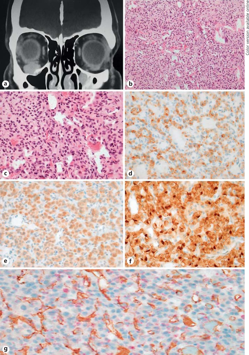
Purpose of the study: To describe the rare occurrence of a paraganglioma in the orbit and how to triage for genetic testing and assess the prognosis with succinate dehydrogenase subunit B (SDHB) immunohistochemical staining.
Method: Case report.
Procedures: A 47-year-old 'healthy' male presented with painless exophthalmos and diplopia secondary to an infraorbital tumour mass.
Results: The orbital biopsy was diagnosed as paraganglioma with positive staining with SDHB.
Conclusion: The rarity of an orbital paraganglioma was followed by the clinical search for a possible occult extraorbital primary paraganglioma. SDHB staining helped in the triage for genetic testing and gave an idea about the prognosis for this tumour.
Keywords: Familial syndrome; Orbit; Paraganglioma; Succinate dehydrogenase immunohistochemical stain.
Figures
References
-
- Sharma MC, Epari S, Gaikwad S, Verma A, Sarkar C. Orbital paraganglioma: report of a rare case. Can J Ophthalmol. 2005;40:640–644. - PubMed
-
- Hill RH, 3rd, Platt SM, Bersani A, Barker-Griffith A, Strumpf K. Regression of a paraganglioma tumor of the orbit. Orbit. 2015;34:99–102. - PubMed
-
- Bagheri A, Aletha M, Salour H, Abdollahi A, Silbert D, Rezaei-Kanavi M. Orbital paraganglioma presenting as lateral rectus enlargement and its novel management: a case report and review of the literature. Orbit. 2012;31:256–260. - PubMed
-
- Makhdoomi R, Nayil K, Santosh V, Kumar S. Orbital paraganglioma: a case report and review of the literature. Clin Neuropatol. 2010;29:100–104. - PubMed
LinkOut - more resources
Full Text Sources
Other Literature Sources
Research Materials
