

SESN2/sestrin 2 induction-mediated autophagy and inhibitory effect of isorhapontigenin (ISO) on human bladder cancers
- PMID: 27171279
- PMCID: PMC4968238
- DOI: 10.1080/15548627.2016.1179403
SESN2/sestrin 2 induction-mediated autophagy and inhibitory effect of isorhapontigenin (ISO) on human bladder cancers
Abstract
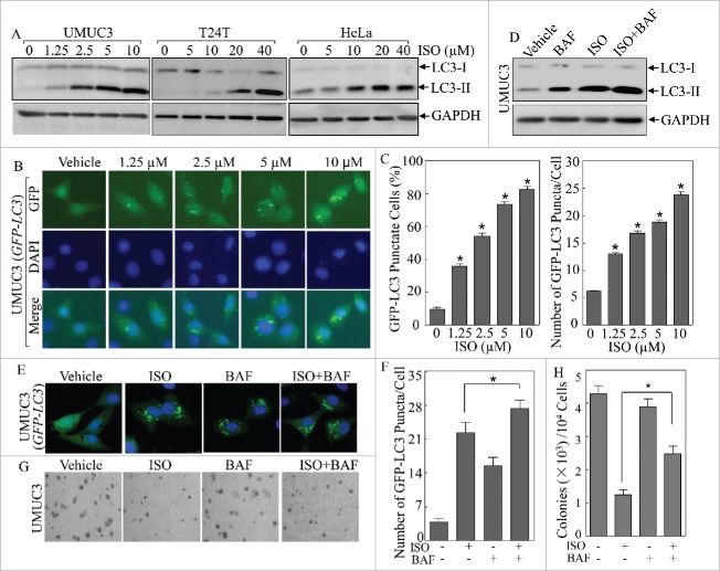
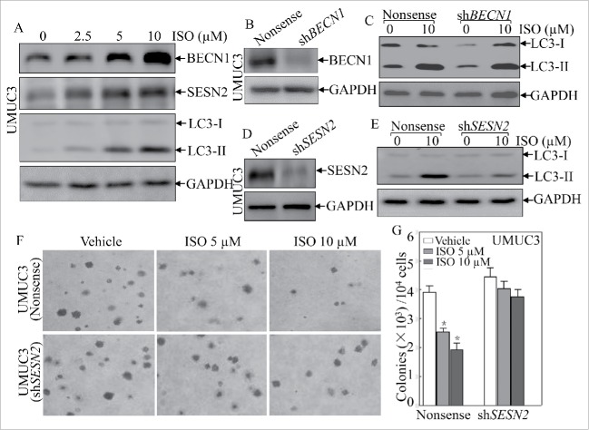
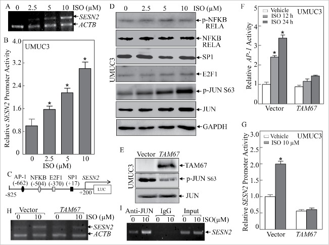
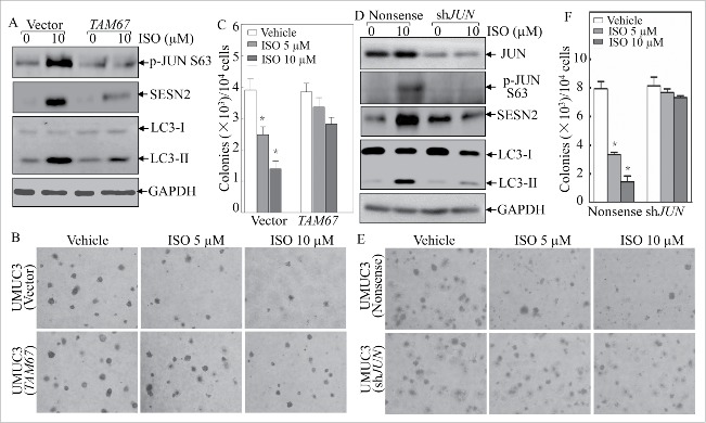
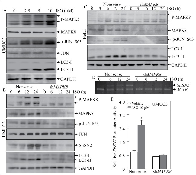
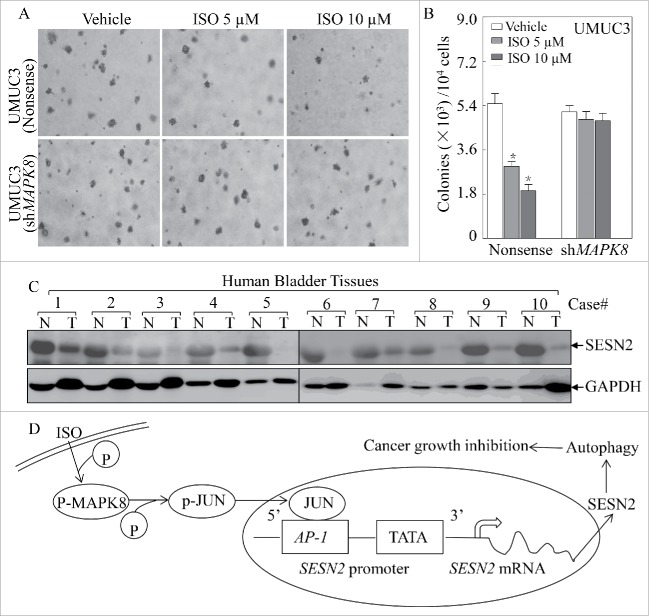
Isorhapontigenin (ISO) is a new derivative of stilbene isolated from the Chinese herb Gnetum cleistostachyum. Our recent studies have revealed that ISO treatment at doses ranging from 20 to 80 μM triggers apoptosis in multiple human cancer cell lines. In the present study, we evaluated the potential effect of ISO on autophagy induction. We found that ISO treatment at sublethal doses induced autophagy effectively in human bladder cancer cells, which contributed to the inhibition of anchorage-independent growth of cancer cells. In addition, our studies revealed that ISO-mediated autophagy induction occurred in a SESN2 (sestrin 2)-dependent and BECN1 (Beclin 1, autophagy related)-independent manner. Furthermore, we identified that ISO treatment induced SESN2 expression via a MAPK8/JNK1 (mitogen-activated protein kinase 8)/JUN-dependent mechanism, in which ISO triggered MAPK8-dependent JUN activation and facilitated the binding of JUN to a consensus AP-1 binding site in the SESN2 promoter region, thereby led to a significant transcriptional induction of SESN2. Importantly, we found that SESN2 expression was dramatically downregulated or even lost in human bladder cancer tissues as compared to their paired adjacent normal tissues. Collectively, our results demonstrate that ISO treatment induces autophagy and inhibits bladder cancer growth through MAPK8-JUN-dependent transcriptional induction of SESN2, which provides a novel mechanistic insight into understanding the inhibitory effect of ISO on bladder cancers and suggests that ISO might act as a promising preventive and/or therapeutic drug against human bladder cancer.
Keywords: MAPK8; autophagy; bladder cancer; isorhapontigenin; sestrin 2.
Figures
Similar articles
-
The Chinese herb isolate isorhapontigenin induces apoptosis in human cancer cells by down-regulating overexpression of antiapoptotic protein XIAP.J Biol Chem. 2012 Oct 12;287(42):35234-35243. doi: 10.1074/jbc.M112.389494. Epub 2012 Aug 15. J Biol Chem. 2012. PMID: 22896709 Free PMC article.
-
HGK-sestrin 2 signaling-mediated autophagy contributes to antitumor efficacy of Tanshinone IIA in human osteosarcoma cells.Cell Death Dis. 2018 Sep 26;9(10):1003. doi: 10.1038/s41419-018-1016-9. Cell Death Dis. 2018. PMID: 30258193 Free PMC article.
-
Cyclin d1 downregulation contributes to anticancer effect of isorhapontigenin on human bladder cancer cells.Mol Cancer Ther. 2013 Aug;12(8):1492-503. doi: 10.1158/1535-7163.MCT-12-0922. Epub 2013 May 30. Mol Cancer Ther. 2013. PMID: 23723126 Free PMC article.
-
Impaired autophagy and APP processing in Alzheimer's disease: The potential role of Beclin 1 interactome.Prog Neurobiol. 2013 Jul-Aug;106-107:33-54. doi: 10.1016/j.pneurobio.2013.06.002. Epub 2013 Jul 1. Prog Neurobiol. 2013. PMID: 23827971 Review.
-
Sestrin2: Its Potential Role and Regulatory Mechanism in Host Immune Response in Diseases.Front Immunol. 2019 Dec 4;10:2797. doi: 10.3389/fimmu.2019.02797. eCollection 2019. Front Immunol. 2019. PMID: 31867002 Free PMC article. Review.
Cited by
-
Induction of RAC1 protein translation and MKK7/JNK-dependent autophagy through dicer/miR-145/SOX2/miR-365a axis contributes to isorhapontigenin (ISO) inhibition of human bladder cancer invasion.Cell Death Dis. 2022 Aug 31;13(8):753. doi: 10.1038/s41419-022-05205-w. Cell Death Dis. 2022. PMID: 36045117 Free PMC article.
-
Carnosic Acid against Lung Cancer: Induction of Autophagy and Activation of Sestrin-2/LKB1/AMPK Signalling.Int J Mol Sci. 2024 Feb 6;25(4):1950. doi: 10.3390/ijms25041950. Int J Mol Sci. 2024. PMID: 38396629 Free PMC article.
-
A paradoxical role for sestrin 2 protein in tumor suppression and tumorigenesis.Cancer Cell Int. 2021 Nov 16;21(1):606. doi: 10.1186/s12935-021-02317-9. Cancer Cell Int. 2021. PMID: 34784907 Free PMC article. Review.
-
Autophagy Regulators in Cancer.Int J Mol Sci. 2023 Jun 30;24(13):10944. doi: 10.3390/ijms241310944. Int J Mol Sci. 2023. PMID: 37446120 Free PMC article. Review.
-
The RING-type E3 ligase RNF186 ubiquitinates Sestrin-2 and thereby controls nutrient sensing.J Biol Chem. 2019 Nov 8;294(45):16527-16534. doi: 10.1074/jbc.AC119.010671. Epub 2019 Oct 4. J Biol Chem. 2019. PMID: 31586034 Free PMC article.
References
-
- Siegel RL, Miller KD, Jemal A. Cancer statistics, 2015. CA: A Cancer J Clin 2015; 65:5-29; http://dx.doi.org/10.3322/caac.21254 - DOI - PubMed
-
- Murphy WM, Soloway MS, Jukkola AF, Crabtree WN, Ford KS. Urinary cytology and bladder cancer. The cellular features of transitional cell neoplasms. Cancer 1984; 53:1555-65; PMID:6697294; http://dx.doi.org/10.1002/1097-0142(19840401)53:7%3c1555::AID-CNCR282053... - DOI - PubMed
-
- Cowan NC, Crew JP. Imaging bladder cancer. Curr Opin Urol 2010; 20:409-13; PMID:20625298; http://dx.doi.org/10.1097/MOU.0b013e32833cbcb9 - DOI - PubMed
-
- von der Maase H, Sengelov L, Roberts JT, Ricci S, Dogliotti L, Oliver T, Moore MJ, Zimmermann A, Arning M. Long-term survival results of a randomized trial comparing gemcitabine plus cisplatin, with methotrexate, vinblastine, doxorubicin, plus cisplatin in patients with bladder cancer. J Clin Oncol: Off J Am Soc Clin Oncol 2005; 23:4602-8; PMID:16034041; http://dx.doi.org/10.1200/JCO.2005.07.757 - DOI - PubMed
-
- Fang Y, Cao Z, Hou Q, Ma C, Yao C, Li J, Wu XR, Huang C. Cyclin d1 downregulation contributes to anticancer effect of isorhapontigenin on human bladder cancer cells. Mol Cancer Ther 2013; 12:1492-503; PMID:23723126; http://dx.doi.org/10.1158/1535-7163.MCT-12-0922 - DOI - PMC - PubMed
Publication types
MeSH terms
Substances
Grants and funding
LinkOut - more resources
Full Text Sources
Other Literature Sources
Medical
Research Materials
Miscellaneous