. 2016 May 12;11(5):e0155359.
doi: 10.1371/journal.pone.0155359. eCollection 2016.

A Novel Method for Gene-Specific Enhancement of Protein Translation by Targeting 5'UTRs of Selected Tumor Suppressors

Affiliations

A Novel Method for Gene-Specific Enhancement of Protein Translation by Targeting 5'UTRs of Selected Tumor Suppressors

Adam Master et al. PLoS One. .

Abstract

Background: Translational control is a mechanism of protein synthesis regulation emerging as an important target for new therapeutics. Naturally occurring microRNAs and synthetic small inhibitory RNAs (siRNAs) are the most recognized regulatory molecules acting via RNA interference. Surprisingly, recent studies have shown that interfering RNAs may also activate gene transcription via the newly discovered phenomenon of small RNA-induced gene activation (RNAa). Thus far, the small activating RNAs (saRNAs) have only been demonstrated as promoter-specific transcriptional activators.

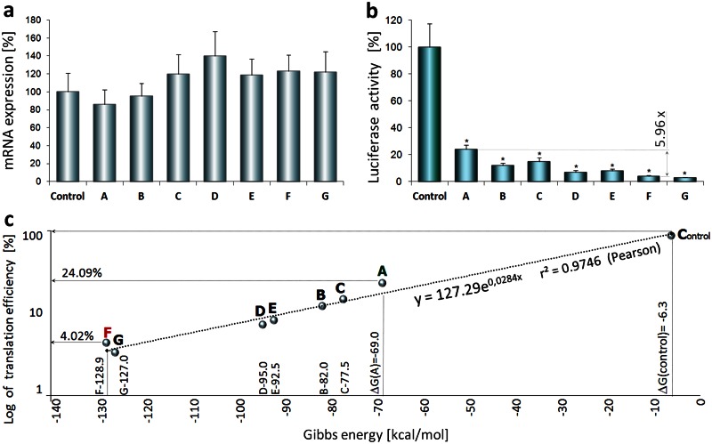
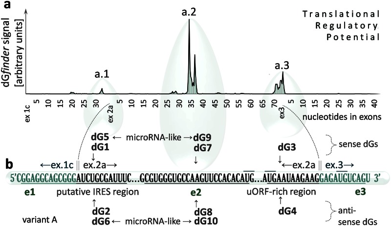
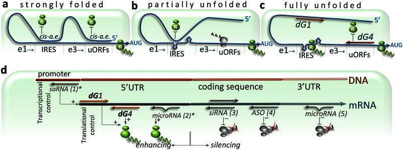
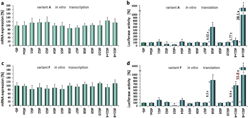
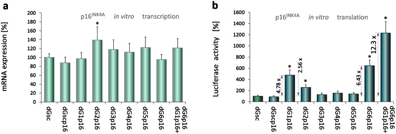
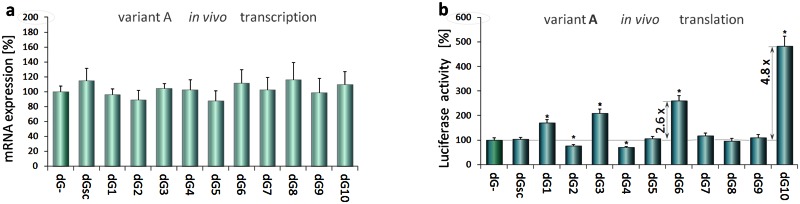
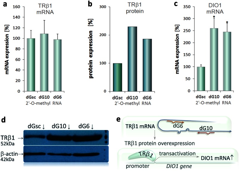
Findings: We demonstrate that oligonucleotide-based trans-acting factors can also specifically enhance gene expression at the level of protein translation by acting at sequence-specific targets within the messenger RNA 5'-untranslated region (5'UTR). We designed a set of short synthetic oligonucleotides (dGoligos), specifically targeting alternatively spliced 5'UTRs in transcripts expressed from the THRB and CDKN2A suppressor genes. The in vitro translation efficiency of reporter constructs containing alternative TRβ1 5'UTRs was increased by up to more than 55-fold following exposure to specific dGoligos. Moreover, we found that the most folded 5'UTR has higher translational regulatory potential when compared to the weakly folded TRβ1 variant. This suggests such a strategy may be especially applied to enhance translation from relatively inactive transcripts containing long 5'UTRs of complex structure.

Significance: This report represents the first method for gene-specific translation enhancement using selective trans-acting factors designed to target specific 5'UTR cis-acting elements. This simple strategy may be developed further to complement other available methods for gene expression regulation including gene silencing. The dGoligo-mediated translation-enhancing approach has the potential to be transferred to increase the translation efficiency of any suitable target gene and may have future application in gene therapy strategies to enhance expression of proteins including tumor suppressors.


Conflict of interest statement

Competing Interests: The authors have declared that no competing interests exist.

Figures

Fig 1
Fig 1. Correlation between Gibbs energy and basic TRβ1 5'UTR-mediated translation efficiency.
(a) Luciferase mRNA levels from in vitro wheat germ-based coupled transcription-translation assay performed on plasmids containing TRβ1 5’UTR variants A-G are shown relative to control plasmid. (b) Effects of 5’UTR variants A-G on luciferase activities after 6h coupled transcription-translation. Three independent experiments were performed in triplicate and shown as mean % mRNA or luciferase activity± SD. Data were analyzed by ANOVA followed by Dunnett’s multiple comparison test; *p< 0.001 vs. control. c, Correlation between the calculated Gibbs energies (X axis) of each 5’UTR variant (S1 Table) and UTR-mediated luciferase translation efficiency. The correlation is shown as the exponential trend-line y = 127.29*e0,0248*X, where x = calculated Gibbs energy; Pearson product-moment correlation coefficient r² = 0,9746. Logarithmically transformed data of translation efficiency (Y axis) were analyzed together with Gibbs energies by linear regression; p = 0.0123.
Fig 2
Fig 2. dGoligo recognition sites.
(a) Cis-acting elements (e1, e2, e3) of variant A of TRβ1 5’UTR determined by dGenhancer, which indicates signal maxima (a.1, a.2 and a.3) corresponding to the 5’UTR fragments with the highest translational regulatory potential. The signal intensity represents transformed mean of 6 consecutive changes in Gibbs energy (ΔG) observed among 5’UTR sequences containing artificial SNPs. The SNPs were used as a theoretical model to calculate which sequence fragments (within the UTR) can change ΔGs (of the UTR) the most, thereby affecting the translational potential of the 5’UTR. (b) dGoligo (dGs 1–10) targets (e1, e2, e3) in TRβ1 5’UTR are shown as underlined sequences. dGs are presented above (sense) and below (antisense) the TRβ1 5’UTR A. Each dG shares homology with the TRβ1 5’UTR, targeting one of the indicated sequences: a putative IRES site on ex1c/ex2a junction (underlined) targeted by dG1, dG2, dG5, dG6, a sequence containing multiple alternate AUGs (uORFs-rich region) and located on ex2a/ex3 junction targeted by dG3, dG4 or a sequence in the middle of exon 2 targeted by dG7 dG8, dG9 and dG10. All dGs were designed as complementary pairs of antisense strand (dG2, dG4, dG6, dG8, dG10) directly recognizing the indicated region and sense strand (dG1, dG3, dG5, dG7, dG9) that can bind to distant sequences that interact through complimentary base-pairing with the indicated region (S1 Fig). Oligonucleotides dG5, dG6, dG9 and dG10 were synthesized as microRNA-like oligonucleotides with 3-nt insertion in the middle of their sequences.
Fig 3
Fig 3. Folding states of TRβ1 5’UTRs.
Translation efficiency of TRβ1 is dependent on folding states of its 5'UTR, which is proposed to be: strongly folded (a), partially unfolded (b) or fully unfolded following interaction with dG1 and dG4 (c). The 5’UTR is shown as curve ended by an arrow at AUG translation start codon. Two linked ovals assigned by letter R represent ribosome that may be blocked by distant cis-acting element (cis-a.e) or trans-acting factor (dG1 or dG4). AUG start codon is preceded by selected translation-regulating elements (e1 and e3). Translation-enhancing element e1 contains putative Internal Ribosome Entry Site (IRES) that may be involved in enhancement of cap-independent translation initiation. Translation-silencing element e3 contains upstream Open Reading Frames (uORFs)–rich region that can reduce translation initiation from correct AUG start codon due to simultaneous synthesis of truncated proteins originated from upstream AUGs (shown by inverted ribosome). a, Theoretical folding state of TRβ1 5'UTR characterized by the presence of highly-structured sequence that can block both: translation-enhancing e1 and translation-silencing e3, finally leading to only basal protein synthesis. (b) Another theoretical folding state described by partially unfolded 5’UTR with blocked e1 and unblocked e3, resulting in basal translation rates as well. (c) Proposed model of dG1 and dG4 -mediated enhancement of translation efficiency, wherein antisense dG4 could lead to repression of uORFs within e3, whereas binding of sense dG1 to a distant sequence (usually folded with e1) could release this translation-enhancing region, allowing for protein over-expression (additional description is given in S4 Fig). (d) dGoligo (dG) targets on mRNA. Locations of dG binding sites are shown in the context of typical targets of the most known small interfering RNAs. microRNA (2, 5), siRNA (3), saRNA (1) and ASO (4), are shown by short grey arrows. Newly described interactions that may result in up-regulation of gene expression are indicated by asterisk*.
Fig 4
Fig 4. dGoligo-mediated gene expression changes under in vitro conditions.
Effects of each DNA oligonucleotides dG1-dG10 (S3 Table), on in vitro transcription of luciferase reporter constructs (panels a, c) and translation efficiency (b, d), using pKS-A (a, b) or pKS-F plasmid (c, d). Data normalized to control (dG-) containing pKS-A or pKS-F without addition of dGoligo. Scrambled control (dGsc) had no effect on transcription or translation. The strongest enhancing effects on luciferase activity were obtained by combining dG1+dG8 (28.10-fold from pKS-A and 55.80-fold from pKS-F). Results from three independent experiments performed in triplicates are shown as mean % mRNA (a, c) or luciferase activity (b, d) ± SD. Data analyzed by ANOVA followed by Dunnett’s multiple comparison test. *p<0.001 vs. control.
Fig 5
Fig 5. dGoligo-mediated upregulation of CDKN2A expression.
PCR-amplified linear luciferase expression construct containing 5’UTR of p16INK4a (CDKN2A) was generated (S1 Appendix) and used as a template in coupled transcription-translation assay that was performed as described in experiments with TRβ1 5'UTRs. (a) Luciferase mRNA levels after 6-hour in vitro reaction of the linear construct with a DNA-based dGoligos dG1p16—dG6p16, dG1p16+dG6p16 or dGscp16 (S3 Table) targeting the p16INK4a 5'UTR. (b) Luciferase activity as a measure of dG-mediated translational control. All data are shown as mean % mRNA (a) or luciferase activity (b) ± SD. Data were analyzed by ANOVA followed by Dunnett’s multiple comparison test. *p< 0.001 vs. control.
Fig 6
Fig 6. dGoligo-mediated gene expression changes in Caki-2 cells.
Effects of 2’-O-methyl RNA oligonucleotides on luciferase transcription (panel a) and translation (b) in Caki-2 cells transfected with pGL3-A. MicroRNA-like dG10 and microRNA-like dG6 exerted the strongest translation-enhancing effects in Caki-2 cells (4.83-fold and 2.60-fold respectively). Results from three independent experiments performed in triplicates are shown as mean % mRNA (a) or luciferase activity(b) ± SD. Data analyzed by ANOVA followed by Dunnett’s multiple comparison test. *p<0.001 vs. control.
Fig 7
Fig 7. Effects of selected dGs on expression of endogenous TRβ1 in Caki-2 cells.
Caki-2 cells were transfected with 2’-O-methyl-modified RNA-based dG6, dG10 or scrambled control—dGsc and cultured (without any plasmid) according to the procedure used in translation-enhancing assay (Materials and Methods). dGs were selected on the basis of previously obtained results (Fig 6). Expression of TRβ1 mRNA (a), protein (b) and DIO1 mRNA (c) are shown in upper panel. Semi-quantitative real-time PCR was performed for TRβ1 (exon 2–3) and DIO1, as described before [23]. Data from three independent experiments were performed in triplicate and shown as mean values ± SD. Statistics were calculated using t-test to compare cells transfected with dGs vs. dGsc. *p<0.001. (d) An example western blot for TRβ1 and β-actin is shown in lower panel. Each band (dGsc, dG10, dG6) represents sample combined from nine protein lysates. Relative density of bands was quantified by densitometry and TRβ1 protein levels were normalized to β-actin. (e) A simplified model of dG-mediated upregulation of endogenous TRβ1 protein, which has been demonstrated before to act as a transcription factor activating transcription of multiple genes including type 1 iodothyronine deiodinase (DIO1).

Similar articles

Cited by

References

    1. Van Der Kelen K, Beyaert R, Inzé D, De Veylder L. Translational control of eukaryotic gene expression. Crit Rev Biochem Mol Biol. 2009. Jul-Aug;44(4):143–68. 10.1080/10409230902882090 - DOI - PubMed
    1. Myasnikov AG, Simonetti A, Marzi S, Klaholz BP. Structure-function insights into prokaryotic and eukaryotic translation initiation. Curr Opin Struct Biol. 2009. June;19(3):300–9. 10.1016/j.sbi.2009.04.010 - DOI - PubMed
    1. Mahen EM, Watson PY, Cottrell JW, Fedor MJ. mRNA secondary structures fold sequentially but exchange rapidly in vivo. PLoS Biol. 2010. February 9;8(2):e1000307 10.1371/journal.pbio.1000307 - DOI - PMC - PubMed
    1. Vattem KM, Wek RC. Reinitiation involving upstream ORFs regulates ATF4 mRNA translation in mammalian cells. Proc Natl Acad Sci U S A. 2004. August 3;101(31):11269–74. - PMC - PubMed
    1. Wellensiek BP, Larsen AC, Stephens B, Kukurba K, Waern K, Briones N, et al. Genome-wide profiling of human cap-independent translation-enhancing elements. Nat Methods. 2013. August;10(8):747–50. 10.1038/nmeth.2522 - DOI - PMC - PubMed

Publication types

MeSH terms

Substances