

Pulmonary Adenocarcinoma Metastatic to the Choroid Diagnosed by Biopsy of an Extrascleral Nodule
- PMID: 27171574
- PMCID: PMC4847665
- DOI: 10.1159/000430098
Pulmonary Adenocarcinoma Metastatic to the Choroid Diagnosed by Biopsy of an Extrascleral Nodule
Abstract
Purpose/background: To report a patient with orbital extension of a choroidal metastasis produced by a pulmonary adenocarcinoma which was diagnosed by biopsy of the extrascleral nodule.
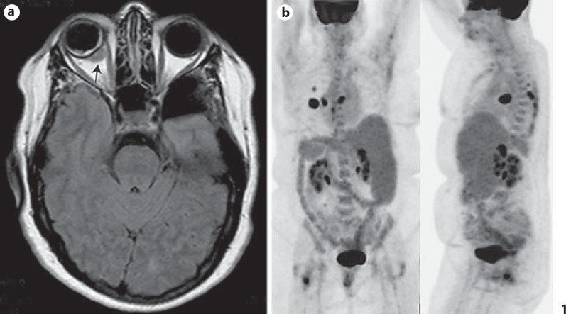
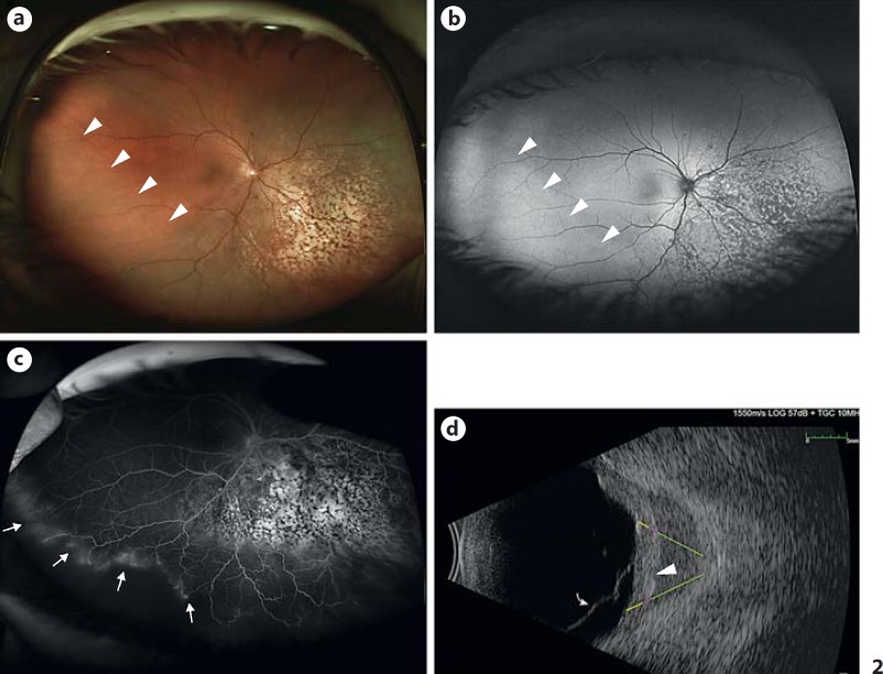
Methods: Clinical history and imaging studies (including fundus photography, autofluorescence, fluorescein angiography, B-scan, and orbital MRI) were reviewed along with histopathologic and immunohistochemical studies.
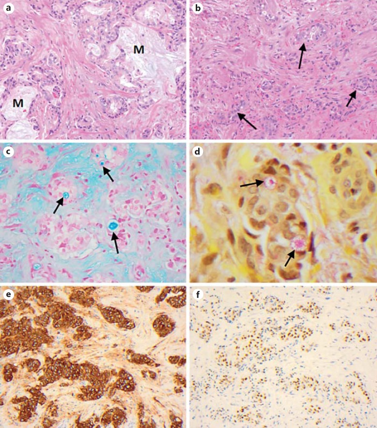
Results: A 60-year-old woman presented with decreased vision in the right eye. Fundus examination revealed a leopard-spotted choroidal lesion and associated serous retinal detachment. Imaging disclosed an enhancing orbital lesion abutting the sclera near the choroidal mass, which had spread outside of the eye. Histopathology revealed lumen-forming cells elaborating mucin. The cells were immunohistochemically positive for epithelial membrane antigen, thyroid transcription factor 1, and cytokeratin 7 and negative for cytokeratin 20. This was consistent with a pulmonary adenocarcinoma. Widespread metastases were subsequently found.
Conclusions: This is the first detailed case report of a successful biopsy of the orbital extension of an essentially posterior intraocular tumor. Such a maneuver permits a much more generous tissue sample than a needle biopsy. In the current case, a large tissue sample provided the basis for complete immunohistochemical evaluation, leading to the diagnosis of an intraocular metastatic mucin-producing adenocarcinoma of lung origin.
Keywords: Choroid; Immunohistochemistry; Lung carcinoma; Metastasis; Orbit.
Figures
Similar articles
-
Papillary thyroid carcinoma: bilateral choroidal metastases with extrascleral extension.Korean J Ophthalmol. 2013 Jun;27(3):215-8. doi: 10.3341/kjo.2013.27.3.215. Epub 2013 Apr 2. Korean J Ophthalmol. 2013. PMID: 23730117 Free PMC article.
-
Small Choroidal Melanoma Revealed by a Large Extrascleral Extension.Ocul Oncol Pathol. 2017 Sep;3(3):240-246. doi: 10.1159/000455870. Epub 2017 Mar 23. Ocul Oncol Pathol. 2017. PMID: 29071276 Free PMC article.
-
Metastatic adenocarcinoma of the cervix presenting as a choroidal mass: A case report and review of literature of cervical metastases to the eye.Indian J Ophthalmol. 2015 Aug;63(8):674-8. doi: 10.4103/0301-4738.169792. Indian J Ophthalmol. 2015. PMID: 26576527 Free PMC article. Review.
-
MULTIMODAL IMAGING AND HISTOLOGIC CORRELATION OF ISOLATED METASTASIS OF PROSTATE ADENOCARCINOMA TO THE CHOROID.Retin Cases Brief Rep. 2017 Spring;11(2):166-170. doi: 10.1097/ICB.0000000000000320. Retin Cases Brief Rep. 2017. PMID: 27124795
-
Mucoepidermoid carcinoma of the conjunctiva with lung metastasis.Indian J Ophthalmol. 2015 May;63(5):457-9. doi: 10.4103/0301-4738.159893. Indian J Ophthalmol. 2015. PMID: 26139812 Free PMC article. Review.
Cited by
-
Choroidal Mucinous Metastatic Adenocarcinoma from the Colon: A Diagnostic Challenge.Ocul Oncol Pathol. 2019 Jan;5(1):66-74. doi: 10.1159/000487598. Epub 2018 Jun 27. Ocul Oncol Pathol. 2019. PMID: 30675480 Free PMC article.
-
From Blurred Vision to Stage IV Lung Cancer: Choroidal Metastases.Cureus. 2025 Jan 22;17(1):e77835. doi: 10.7759/cureus.77835. eCollection 2025 Jan. Cureus. 2025. PMID: 39991411 Free PMC article.
-
Sclerosing Signet Ring Cell Carcinoma of the Lacrimal Gland: A Potentially New Primary Entity.Ocul Oncol Pathol. 2020 Aug;6(4):265-274. doi: 10.1159/000505490. Epub 2020 Feb 7. Ocul Oncol Pathol. 2020. PMID: 33005616 Free PMC article.
References
-
- Ferry AP, Font RL. Carcinoma metastatic to the eye and orbit. 1. A clinicopathologic study of 227 cases. Arch Ophthalmol. 1974;92:276–286. - PubMed
-
- Shields CL, Shields JA, Gross NE, et al. Survey of 520 eyes with uveal metastases. Ophthalmology. 1997;104:1265–1276. - PubMed
-
- Abundo RE, Orenic CJ, Anderson SF, et al. Choroidal metastases resulting from carcinoma of the lung. J Am Optom Assoc. 1997;68:95–108. - PubMed
-
- Bianciotto C, Demirci H, Shields CL, et al. Metastatic tumors to the eyelid: report of 20 cases and review of the literature. Arch Ophthalmol. 2009;127:999–1005. - PubMed
Grants and funding
LinkOut - more resources
Full Text Sources
Other Literature Sources
Research Materials
