. 2016 Jul 2;12(7):1168-79.
doi: 10.1080/15548627.2016.1179402. Epub 2016 May 12.

SLC35D3 increases autophagic activity in midbrain dopaminergic neurons by enhancing BECN1-ATG14-PIK3C3 complex formation

Affiliations

SLC35D3 increases autophagic activity in midbrain dopaminergic neurons by enhancing BECN1-ATG14-PIK3C3 complex formation

Zong-Bo Wei et al. Autophagy. .

Abstract

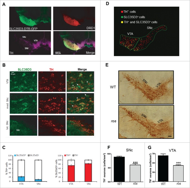
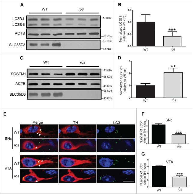
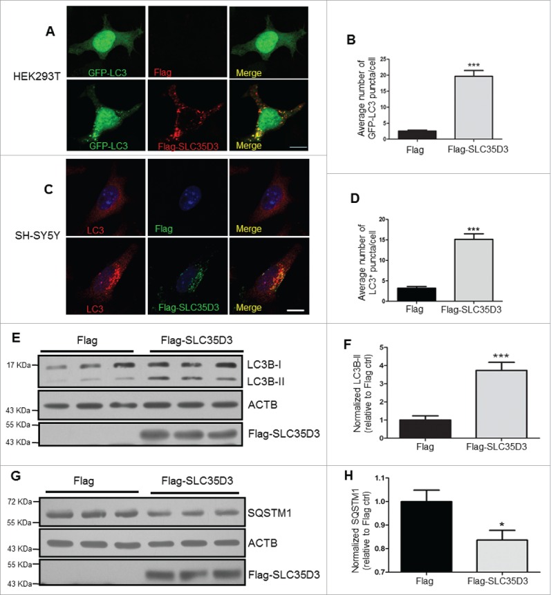
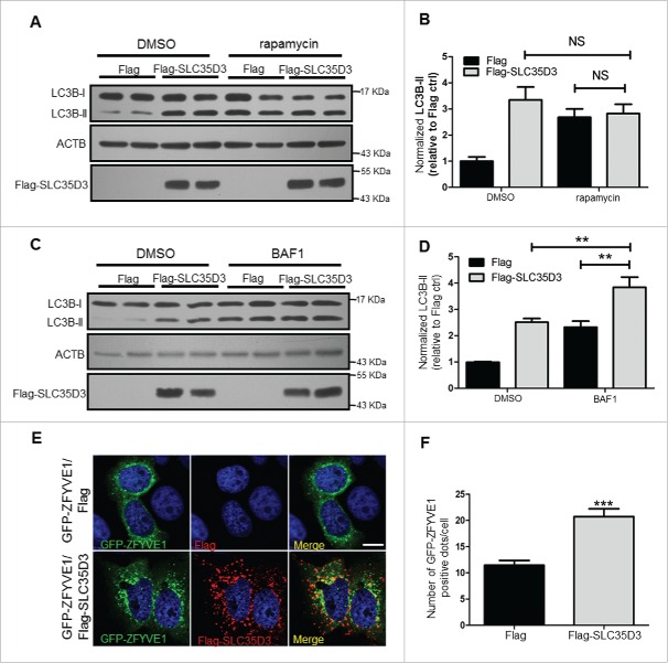
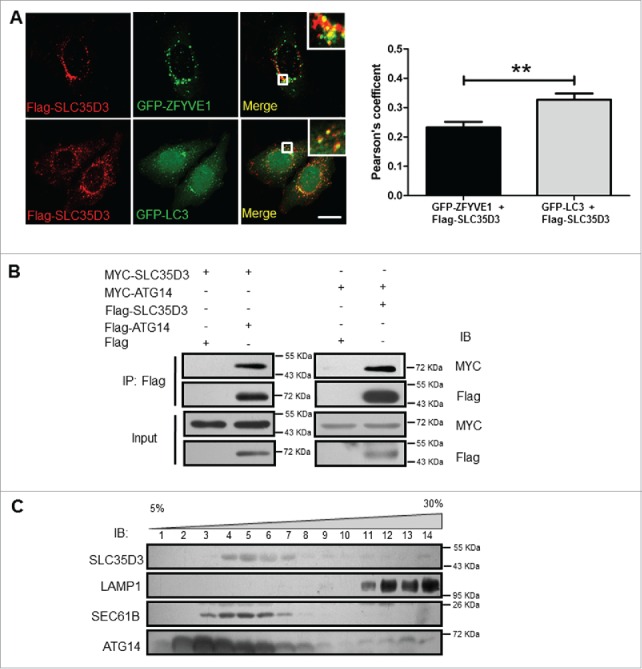
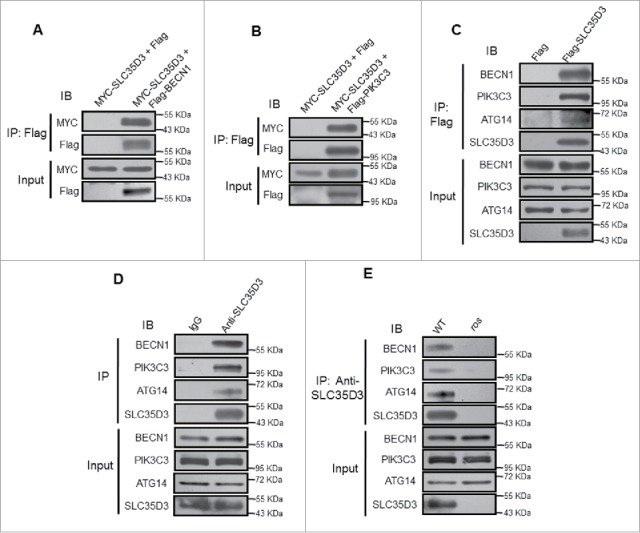
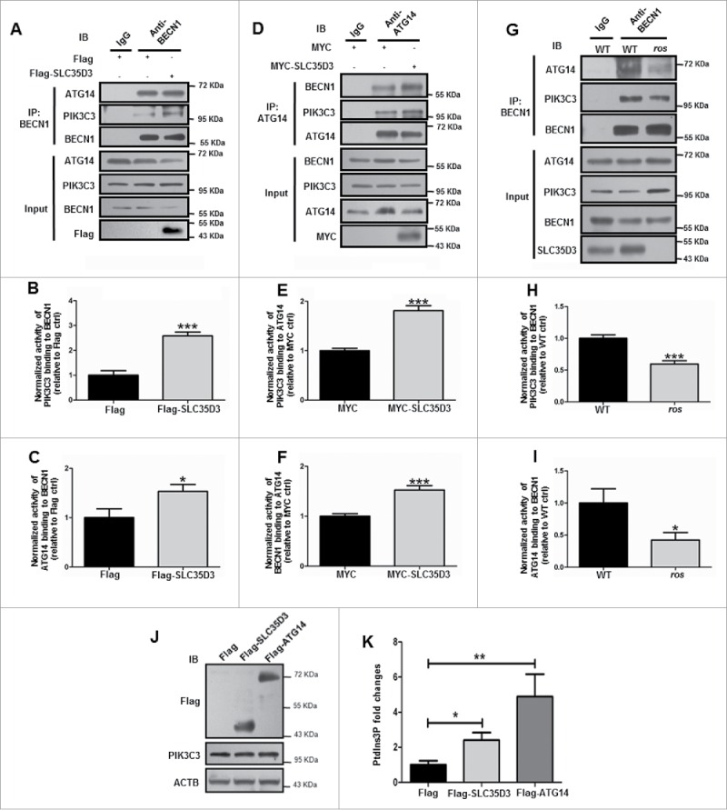
Searching for new regulators of autophagy involved in selective dopaminergic (DA) neuron loss is a hallmark in the pathogenesis of Parkinson disease (PD). We here report that an endoplasmic reticulum (ER)-associated transmembrane protein SLC35D3 is selectively expressed in subsets of midbrain DA neurons in about 10% TH (tyrosine hydroxylase)-positive neurons in the substantia nigra pars compacta (SNc) and in about 22% TH-positive neurons in the ventral tegmental area (VTA). Loss of SLC35D3 in ros (roswell mutant) mice showed a reduction of 11.9% DA neurons in the SNc and 15.5% DA neuron loss in the VTA with impaired autophagy. We determined that SLC35D3 enhanced the formation of the BECN1-ATG14-PIK3C3 complex to induce autophagy. These results suggest that SLC35D3 is a new regulator of tissue-specific autophagy and plays an important role in the increased autophagic activity required for the survival of subsets of DA neurons.

Keywords: BECN1-ATG14-PIK3C3 complex; Parkinson disease; SLC35D3; autophagy; dopaminergic neuron; neurodegeneration.


Figures

Figure 1.
Figure 1.
SLC35D3 is differentially expressed in subsets of DA neurons in the midbrain and the ros mice show reduced TH+ neurons. (A) In our DTR-GFP transgenic mice (3 month old, 2 males), the Slc35d3-promoter driven GFP signals were present in the projections of striatal DRD1-positive neurons in the substantia nigra pars reticutala (SNr), and in the cell bodies of the TH-positive neurons located in the substantia nigra pars compacta (SNc) and in the ventral tegmental area (VTA). (B) Colabeling of SLC35D3-GFP (green) and TH (red) in the cell bodies of the neurons located in the VTA, medial SNc and lateral SNc. Note that only a portion of TH+ neurons express SLC35D3 (asterisk), some neurons express SLC35D3 but not TH (arrow), some neurons express TH but not SLC35D3. (C) In the SNc, 9.46 ± 2.08 % TH+ cells was SLC35D3+, and 83.12 ± 2.21 % SLC35D3+ cells was TH+. By contrast, in the VTA, 22.06 ± 3.35 % TH+ cells was SLC35D3+, and 74.06 ± 5.10 % SLC35D3+ cells was TH+. The quantifications were performed on 2 mice and the number of cells was counted on 40-µm thick section (n=8 sections per mouse, both hemispheres). (D) Schematic distribution pattern of different population neurons in the midbrain (right hemisphere) from the images in (B). Note that in the SNc, SLC35D3+ cells were almost exclusively located in the median SNc compared to the lateral part. (E) Immunohistochemical detection of TH on the coronal brain sections of the midbrain at 6 mo of age in both wild-type (WT) and ros mice. Scale bar: 100 µm. (F) Ros mice showed a 11.9 % reduction in cell density of TH+ neurons in the SNc. WT: 956 ± 11 cells/mm2; ros: 842 ± 19 cells/mm2, n = 5; ***, P < 0.001. (G) Ros mice showed a 15.5% reduction in cell density of TH+ neurons in the VTA. WT: 679 ± 19 cells/mm2, ros, 574 ± 8 cells/mm2, n = 5; ***, P < 0.001.
Figure 2.
Figure 2.
Reduced LC3B-II levels in ros midbrain. (A, B) Steady-state level of LC3B-II in the midbrain containing the SN and VTA was lower in ros mice compared with wild type (WT). ACTB/β-actin was used as a loading control. Bars represent mean ± SEM, WT: n = 12, ros: n=11; ***, P < 0.001. (C, D) Steady-state level of insoluble SQSTM1 in the SN and VTA was increased in ros mice compared with WT mice. ACTB was used as a loading control. Bars represent mean ± SEM, n = 3; **, P < 0.01. (E) Representative images of LC3 puncta (green) stained with anti-LC3 antibody in TH-labeled (red) neurons. Arrows show the LC3 -positive dots. Scale bar: 10 μm. (F) The number of autophagosomes in TH+ neurons from the SNc was reduced in ros mice. 225 neurons were counted from 5 WT mice, 238 neurons were counted from 5 ros mice. WT, 6.40 ± 0.39 dots/TH+ neuron; ros, 3.15 ± 0.29 dots/TH+ neuron; ***, P < 0.001. (G) The number of autophagosomes in TH+ neurons from the VTA was reduced in ros mice. 239 neurons were counted from 5 WT mice, 261 neurons were counted from 5 ros mice. WT, 6.10 ± 0.27 dots/TH+ neuron; ros, 2.95 ± 0.34 dots/TH+ neuron; ***, P < 0.001.
Figure 3.
Figure 3.
Expression of SLC35D3 increases autophagic activity. (A, B) In HEK293T cells, cotransfection of GFP-LC3 and Flag-SLC35D3 increased LC3 puncta. Scale bar: 10 µm; for statistics, at least 50 cells were counted; ***; P < 0.001. (C, D) In SH-SY5Y cells, transfection of Flag-SLC35D3 increased LC3 puncta. Scale bar: 10 µm; for statistics, at least 50 cells were counted; ***, P < 0.001. (E, F) Steady-state level of LC3B-II in the SLC35D3-transfected cells was increased compared to the control cells. ACTB was used as a loading control. Bars represent mean ± SEM, n = 11; ***, P < 0.001. (G, H) Steady-state level of insoluble SQSTM1 in the SLC35D3-transfected cells was decreased compared to the control cells. ACTB was used as loading control. Bars represent mean ± SEM, n = 5; *, P < 0.05.
Figure 4.
Figure 4.
SLC35D3 functions in the early stage of autophagic flux. (A, B) Immunoblotting of LC3B-II in the HEK293T cells transfected with Flag or Flag-SLC35D3 plasmid and treated with DMSO or rapamycin (100 nM) for 4 h. ACTB was used as a loading control. There were no significant changes (N.S., P > 0.05) of LC3B-II after rapamycin treatment. Bars represent mean ± SEM, n = 6. (C, D) Immunoblotting of LC3B-II in the HEK293T cells transfected with Flag or Flag-SLC35D3 plasmid and treated with DMSO or BAF1 (400 nM) for 4 h. ACTB was used as a loading control. The increase of LC3B-II by SLC35D3 expression was further enhanced after BAF1 treatment. Bars represent mean ± SEM, n = 6. **, P < 0.01. (E, F) Expression of SLC35D3 leads to an increase of omegasomes in the HeLa cell. Representative images of GFP-ZFYVE1 puncta in HeLa cells cotransfected with GFP-ZFYVE1 and Flag or GFP-ZFYVE1 and Flag-SLC35D3 plasmids. Scale bar, 10 μm. Quantitative analysis of GFP-ZFYVE1 puncta in the transfected cells showed an increase of GFP-ZFYVE1 dots when cotransfected with Flag-SLC35D3 (55 cells were counted; ***, P < 0.001).
Figure 5.
Figure 5.
SLC35D3 is localized to the autophagy initiation site. (A) Partial colocalization of SLC35D3 with ZFYVE1 or GFP-LC3. HeLa cells were cotransfected with GFP-ZFYVE1/Flag-SLC35D3 or GFP-LC3/Flag-SLC35D3. Cells were fixed and permeabilized after 18 h. SLC35D3 (red) partially colocalizes with the ZFYVE1 or LC3 (green). Insets showed the enlarged white rectangle area. Scale bar: 10 µm. Quantification of colocalization represented by the Pearson correlation coefficient showed that Flag-SLC35D3 colocalized more with GFP-LC3 than with GFP-ZFYVE1. **, P < 0.01, n = 50 cells. (B) Interactions between SLC35D3 and ATG14. CoIP assays showed that Flag-ATG14 coprecipitated with MYC-SLC35D3 (left panel), and Flag-SLC35D3 coprecipitated with MYC-ATG14 (right panel). The Flag-empty vector was used as a negative control. (C) OptiPrep (5–30% [w/v]) gradient assay showed that SLC35D3 coexisted with the ER marker SEC61B, but segregated with the lysosome marker LAMP1. ATG14 had a broader distribution overlapping with SLC35D3 and the lysosomal fractions. Fractions 1 and 14 correspond to the top and bottom ends of the gradient, respectively. IB, immunoblotting; IP, immunoprecipitation.
Figure 6.
Figure 6.
SLC35D3 interacts with the BECN1-ATG14-PIK3C3 complex. (A) Coimmunoprecipitation (coIP) assays showed that Flag-BECN1 coprecipitated with MYC-SLC35D3. The Flag-empty vector was used as a negative control. (B) coIP assays showed that Flag-PIK3C3 coprecipitated with MYC-SLC35D3. The Flag-empty vector was used as a negative control. (C) Affinity isolation assays showed that Flag-SLC35D3 pulled down BECN1, PIK3C3 and ATG14. The Flag-empty vector was used as a negative control. (D) Endogenous IP assays of brain lysates showed that SLC35D3 precipitated BECN1, PIK3C3 and ATG14. IgG was used as a negative control. (E) Endogenous IP assays of brain lysates showed that SLC35D3 precipitated BECN1, PIK3C3 and ATG14 in wild-type brain but not in ros brain. All the above IP assays were repeated independently at least 3 times. IB, immunoblotting; IP, immunoprecipitation.
Figure 7.
Figure 7.
Expression of SLC35D3 enhances the BECN1-ATG14-PIK3C3 complex formation. (A) Expression of Flag-SLC35D3 enhanced BECN1-PIK3C3 and BECN1-ATG14 interactions in HEK293T cells. (B, C) The quantification data of 3 independent experiments in (A) are shown. Bars represent mean ± SEM, n = 3. *, P < 0.05; ***, P < 0.001. (D) Expression of MYC-SLC35D3 increased ATG14-BECN1 and ATG14-PIK3C3 interactions in HKE293T cells. (E, F) The quantification data of 3 independent experiments in (D) are shown. Bars represent mean ± SEM, n = 3. ***, P < 0.001. (G) Reduced BECN1-PIK3C3 and BECN1-ATG14 interactions in the SN and VTA of 6-mo-old ros mice. (H, I) The quantification data of 3 independent experiments in (G) are shown. Bars represent mean ± SEM, n = 3. **, P < 0.01; ***, P < 0.001. (J) PIK3C3 activity was detected by analyzing PtdIns3P production using ELISA assay as described in Materials and Methods. The expression of indicated proteins was verified by immunoblotting. (K) The PtdIns3P fold changes were measured based on the concentration of PtdIns3P and normalized to the Flag control group. Both the expression of Flag-SLC35D3 and Flag-ATG14 enhanced the PIK3C3 activities. Bars represent mean ± SEM, n = 9; *, P < 0.05; **, P < 0.01. IP: immunoprecipitation.

References

    1. Hara T, Nakamura K, Matsui M, Yamamoto A, Nakahara Y, Suzuki-Migishima R, Yokoyama M, Mishima K, Saito I, Okano H, et al.. Suppression of basal autophagy in neural cells causes neurodegenerative disease in mice. Nature 2006; 441:885-9; PMID:16625204; http://dx.doi.org/10.1038/nature04724 - DOI - PubMed
    1. Komatsu M, Waguri S, Chiba T, Murata S, Iwata J, Tanida I, Ueno T, Koike M, Uchiyama Y, Kominami E, et al.. Loss of autophagy in the central nervous system causes neurodegeneration in mice. Nature 2006; 441:880-4; PMID:16625205; http://dx.doi.org/10.1038/nature04723 - DOI - PubMed
    1. Yuan Y, Wang H, Wei Z, Li W. Impaired autophagy in hilar mossy cells of the dentate gyrus and its implication in schizophrenia. J Genet Genomics 2015; 42:1-8; PMID:25619597; http://dx.doi.org/10.1016/j.jgg.2014.12.001 - DOI - PubMed
    1. Nixon RA. The role of autophagy in neurodegenerative disease. Nat Med 2013; 19:983-97; PMID:23921753; http://dx.doi.org/10.1038/nm.3232 - DOI - PubMed
    1. Funke C, Schneider SA, Berg D, Kell DB. Genetics and iron in the systems biology of Parkinson's disease and some related disorders. Neurochem Int 2013; 62:637-52; PMID:23220386; http://dx.doi.org/10.1016/j.neuint.2012.11.015 - DOI - PubMed

MeSH terms