

Multimodal Imaging of Spontaneously Shifting Primary Vitreoretinal Lymphoma
- PMID: 27172327
- PMCID: PMC4847661
- DOI: 10.1159/000374121
Multimodal Imaging of Spontaneously Shifting Primary Vitreoretinal Lymphoma
Abstract
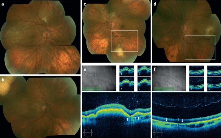
Purpose: To correlate spectral domain optical coherence tomography (SD-OCT) and photographic imaging before and after spontaneous regression of primary vitreoretinal lymphoma (PVRL) lesions.
Procedures: We report the case of a 60-year-old female.
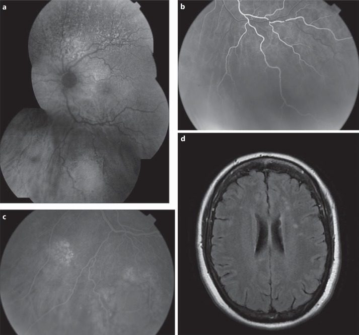
Results: The patient presented with bilateral creamy deposits under the retina and retinal pigment epithelium (RPE), and lesions were visible along Bruch's membrane with SD-OCT and suspicious for PVRL. Systemic workup revealed nonspecific areas of enhancement on neuroimaging. The patient was largely asymptomatic and the decision was made to observe her. Three months later, a new lesion pattern had developed. The color fundus photographs and SD-OCT demonstrated spontaneous regression of the largest sub-RPE lesion, leaving areas of RPE atrophy, while a new larger sub-RPE lesion had formed in the other eye. Vitreous biopsy showed lymphocytes and no malignant cells, while sub-RPE biopsy of the newly formed lesion revealed highly atypical cells positive for CD19 and CD20.
Conclusions: Multimodal imaging documents that PVRL lesion regression and early RPE changes can develop within a 3-month period. Immune control is an important factor in lesion regression in the eye.
Keywords: Immune surveillance; Primary vitreoretinal lymphoma; Spectral domain optical coherence tomography; Spontaneous regression.
Figures
Similar articles
-
Multimodal diagnostic imaging in primary vitreoretinal lymphoma.Int J Retina Vitreous. 2022 Aug 26;8(1):58. doi: 10.1186/s40942-022-00405-0. Int J Retina Vitreous. 2022. PMID: 36028905 Free PMC article. Review.
-
Characteristic optical coherence tomography findings in patients with primary vitreoretinal lymphoma: a novel aid to early diagnosis.Br J Ophthalmol. 2018 Oct;102(10):1362-1366. doi: 10.1136/bjophthalmol-2017-311612. Epub 2018 Jan 6. Br J Ophthalmol. 2018. PMID: 29306864
-
Two cases of primary vitreoretinal lymphoma: a diagnostic challenge : The supporting role of multimodal imaging in the diagnosis of primary vitreoretinal lymphoma.Int Ophthalmol. 2018 Feb;38(1):353-361. doi: 10.1007/s10792-016-0422-1. Epub 2016 Dec 30. Int Ophthalmol. 2018. PMID: 28039672
-
Bilateral primary vitreoretinal lymphoma masquerading as fungal endophthalmitis- a case report.J Ophthalmic Inflamm Infect. 2024 Dec 30;14(1):69. doi: 10.1186/s12348-024-00426-w. J Ophthalmic Inflamm Infect. 2024. PMID: 39738772 Free PMC article.
-
Recent progress in the diagnosis and treatment of primary vitreoretinal lymphoma.Taiwan J Ophthalmol. 2016 Oct-Dec;6(4):170-176. doi: 10.1016/j.tjo.2016.05.002. Epub 2016 Jun 27. Taiwan J Ophthalmol. 2016. PMID: 29018736 Free PMC article. Review.
Cited by
-
Retromode imaging in vitreoretinal lymphoma.Graefes Arch Clin Exp Ophthalmol. 2025 May;263(5):1451-1459. doi: 10.1007/s00417-024-06703-7. Epub 2024 Dec 8. Graefes Arch Clin Exp Ophthalmol. 2025. PMID: 39645624
-
Optical Coherence Tomography Benefits the Diagnosis and Follow-Up of Primary Central Nervous System Lymphoma with Intraocular Involvement.Cancer Manag Res. 2022 Mar 5;14:1007-1018. doi: 10.2147/CMAR.S353142. eCollection 2022. Cancer Manag Res. 2022. PMID: 35283643 Free PMC article.
-
Vitreoretinal Lymphoma.Cancers (Basel). 2021 Aug 4;13(16):3921. doi: 10.3390/cancers13163921. Cancers (Basel). 2021. PMID: 34439078 Free PMC article. Review.
-
Secondary vitreoretinal lymphoma with spontaneous regression.Am J Ophthalmol Case Rep. 2022 Jan 26;25:101357. doi: 10.1016/j.ajoc.2022.101357. eCollection 2022 Mar. Am J Ophthalmol Case Rep. 2022. PMID: 35146209 Free PMC article.
-
Multimodal diagnostic imaging in primary vitreoretinal lymphoma.Int J Retina Vitreous. 2022 Aug 26;8(1):58. doi: 10.1186/s40942-022-00405-0. Int J Retina Vitreous. 2022. PMID: 36028905 Free PMC article. Review.
References
-
- Chan CC, Rubenstein JL, Coupland SE, Davis JL, Harbour JW, Johnston PB, Cassoux N, Touitou V, Smith JR, Batchelor TT, Pulido JS. Primary vitreoretinal lymphoma: a report from an International Primary Central Nervous System Lymphoma Collaborative Group symposium. Oncologist. 2011;16:1589–1599. - PMC - PubMed
-
- Kase S, Namba K, Jin XH, Kubota KC, Ishida S. Spontaneous regression of intraocular lymphoma. Ophthalmology. 2012;119:1083–1084. - PubMed
-
- Iihara K, Yamaguchi K, Nishimura Y, Iwasaki T, Suzuki K, Hirabayashi Y. Spontaneous regression of malignant lymphoma of the breast. Pathol Int. 2004;54:537–542. - PubMed
-
- Ono K. Clinical significance of natural killing activity in patients with advanced lymphoma. J Clin Immunol. 1998;18:132–141. - PubMed
Publication types
Grants and funding
LinkOut - more resources
Full Text Sources
Other Literature Sources
