

Use of a semi-automated cardiac segmentation tool improves reproducibility and speed of segmentation of contaminated right heart magnetic resonance angiography
- PMID: 27173489
- PMCID: PMC5562952
- DOI: 10.1007/s10554-016-0906-0
Use of a semi-automated cardiac segmentation tool improves reproducibility and speed of segmentation of contaminated right heart magnetic resonance angiography
Abstract
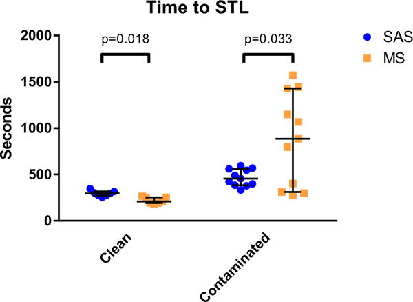
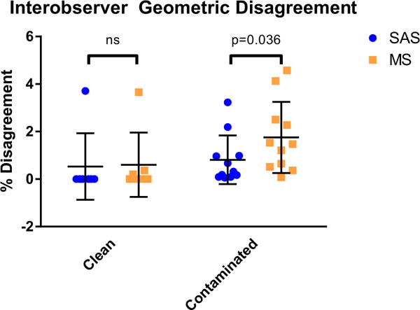
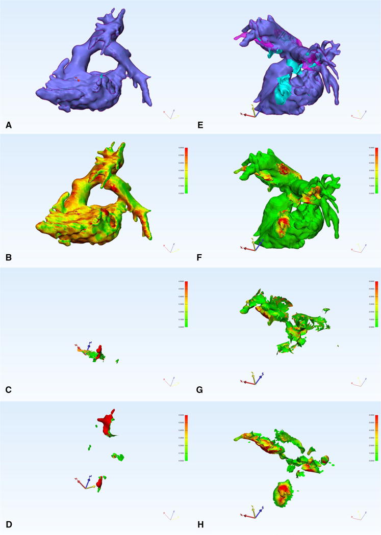
Three-dimensional printing has an increasing number of clinical applications in pediatric cardiology. Time required for dataset segmentation and conversion to stereolithography (STL) format remains a significant limitation. We investigated the impact of semi-automated cardiovascular-specific segmentation software on time and reproducibility of segmentation. Magnetic resonance angiograms (MRAs) of 19 patients undergoing intervention for right ventricular outflow lesions were segmented to demonstrate the right heart. STLs were created by two independent clinicians using semi-automated cardiovascular segmentation (SAS) and traditional manual segmentation (MS). Time was recorded and geometric STL disagreement was determined (0 % = no disagreement, 100 % = complete disagreement). MRA datasets were categorized as clean when only right heart structures were present in the MRA, or contaminated when left heart structures were also present and required removal. Eighteen (seven clean and 11 contaminated) cases were successfully segmented with both methods. Time to STL for clean datasets was faster with MS than SAS [median 209 s (IQR 192-252) vs. 296 s (272-317), p = 0.018] while contaminated datasets were faster with SAS [455 s (384-561) vs. 866 s (310-1429), p = 0.033]. Interobserver STL geometric disagreement was significantly lower using SAS than MS overall (0.70 ± 1.15 % vs. 1.31 ± 1.52 %, p = 0.030), and for the contaminated subset (0.81 ± 1.08 % vs. 1.75 ± 1.57 %, p = 0.036). Most geometric disagreement occurred at areas where left heart contamination was removed. Semi-automated segmentation was faster and more reproducible for contaminated datasets, while MS was faster but equally reproducible for clean datasets. Semi-automated segmentation methods are preferable for contaminated datasets and continued refinement of these tools should be supported.
Keywords: 3D printing; Cardiac segmentation; Magnetic resonance angiography; Stereolithography.
Conflict of interest statement
All authors report no conflict of interest. AT, AKD, JMD, GFG, and TH all work at Children’s Medical Center, Dallas. We have full control of the primary data and agree to allow review of the data if requested.
Figures
References
-
- Dankowski R, Baszko A, Sutherland M, Firek L, Kalmucki P, Wroblewska K, Szyszka A, Groothuis A, Siminiak T. 3D heart model printing for preparation of percutaneous structural interventions: description of the technology and case report. Kardiol Pol. 2014;6:546–551. doi: 10.5603/KP.2014.0119. - DOI - PubMed
MeSH terms
Substances
Grants and funding
LinkOut - more resources
Full Text Sources
Other Literature Sources
