Review
. 2016 Jul;469(1):3-17.
doi: 10.1007/s00428-016-1945-6. Epub 2016 May 12.

Lymph vessels: the forgotten second circulation in health and disease

Affiliations
Review

Lymph vessels: the forgotten second circulation in health and disease

Lukasz A Adamczyk et al. Virchows Arch. 2016 Jul.

Abstract

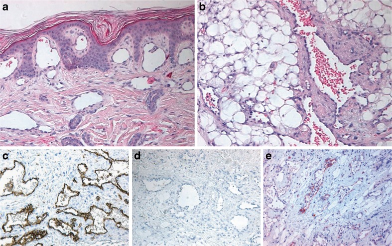
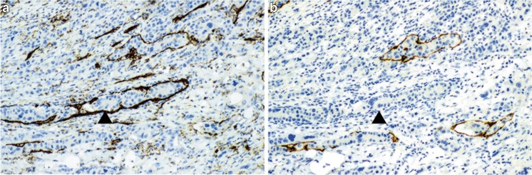
The lymphatic circulation is still a somewhat forgotten part of the circulatory system. Despite this, novel insights in lymph angiogenesis in health and disease, application of immune markers for lymphatic growth and differentiation and also the introduction of new imaging techniques to visualize the lymphatic circulation have improved our understanding of lymphatic function in both health and disease, especially in the last decade. These achievements yield better understanding of the various manifestations of lymph oedemas and malformations, and also the patterns of lymphovascular spread of cancers. Immune markers that recognize lymphatic endothelium antigens, such as podoplanin, LYVE-1 and Prox-1, can be successfully applied in diagnostic pathology and have revealed (at least partial) lymphatic differentiation in many types of vascular lesions.

Keywords: Angiogenesis; Atherosclerosis; Circulation; Genetics; Immunohistochemistry; Lymph vessels; Lymphedema; Metastasis; Vascular malformation; Vascular pathology.


Figures

Fig. 1
Fig. 1
Intravital microscopy photographs showing a lymphatic vessel in diastole (a) and systole (b). The white arrows mark the vessel wall and the black arrow marks the valve. The corresponding diameter changes are depicted in (c). Adapted with permission from Am J Physiol Heart Circ Physiol 2007;293:H709-18
Fig. 2
Fig. 2
Lymphoscintigraphy of a healthy subject (a). Depots at the injection sites on the feet and lymphatic vessels draining the injections sites can be seen as well as the lymph nodes in the groin that the vessels lead to. Lymphatic failure in a patient with Milroy’s disease (b). No lymphatic vessels or uptake in the groin can be seen. Lymphoscintigraphy of a patient with lymphedema distichiasis syndrome (c). There is dermal reflux of lymph fluid in the lower legs. No clear vessels can be seen and there is reduced uptake in the lymph nodes in the groin
Fig. 3
Fig. 3
Classification pathway for primary lymphedema. The pathway provides an overview over the different primary lymphedemas, clinical features and causal genes. Fh = family history. Adapted with permission from Clinical Genetics 2013;84:303–314
Fig. 4
Fig. 4
Lymphatic malformation with intralesional formation of lymph follicles (ac); Haematoxylin and eosin stain (HE) (a); anti-CD31 immunostain (b); anti-D2-40 immunostain of endothelium (c); macrocystic lymphatic malformation (de): HE (d); CD31, low intensity to absent immunostaining with D2-40 antibody of endothelial cells at cystic structures (e) (compared with D2-140 immunostain in (f)); Traumatic changes and haemorrhage in LM (gh), with hobnail-type endothelium in (h). D2-40 immunostaining of microvessels (i)
Fig. 5
Fig. 5
Multiple lymphangioendotheliomatosis. Skin surface with subepidermal dilated thin-walled vessels (HE) (a); deep-seated atypical vessels in subcutis, partially filled with erythrocytes (HE) (b); CD31 immunostain of atypical microvessels (c); absent D2-40 immunostaining (d); focal LYVE-1 immunostaining of microvessels (e)
Fig. 6
Fig. 6
Detail of tumour (melanoma) with lymphovascular invasion; CD31 immunostain showing immunoreactivity of endothelium in all microvessels (a); D2-40 immunostain showing immunoreactivity of endothelium with intravascular tumour deposit (b)

References

    1. Baluk P, Fuxe J, Hashizume H, Romano T, Lashnits E, Butz S, Vestweber D, Corada M, Molendini C, Dejana E, McDonald DM. Functionally specialized junctions between endothelial cells of lymphatic vessels. J Exp Med. 2007;204:2349–2362. doi: 10.1084/jem.20062596. - DOI - PMC - PubMed
    1. Briggs Boedtkjer D, Rumessen J, Baandrup U, Skov Mikkelsen M, Telinius N, Pilegaard H, Aalkjaer C, Hjortdal V. Identification of interstitial Cajal-like cells in the human thoracic duct. Cells Tissues Organs. 2013;197:145–158. doi: 10.1159/000342437. - DOI - PubMed
    1. McCloskey KD, Hollywood MA, Thornbury KD, Ward SM, McHale NG. Kit-like immunopositive cells in sheep mesenteric lymphatic vessels. Cell Tissue Res. 2002;310:77–84. doi: 10.1007/s00441-002-0623-y. - DOI - PubMed
    1. Renkin EM. Some consequences of capillary permeability to macromolecules: starling’s hypothesis reconsidered. Am J Physiol. 1986;250:H706–H710. - PubMed
    1. Levick JR, Michel CC. Microvascular fluid exchange and the revised Starling principle. Cardiovasc Res. 2010;87:198–210. doi: 10.1093/cvr/cvq062. - DOI - PubMed

MeSH terms

LinkOut - more resources