. 2016 May 11;19(5):686-95.
doi: 10.1016/j.chom.2016.04.005.

HIV-1 Vpu Mediates HLA-C Downregulation

Affiliations

HIV-1 Vpu Mediates HLA-C Downregulation

Richard Apps et al. Cell Host Microbe. .

Abstract

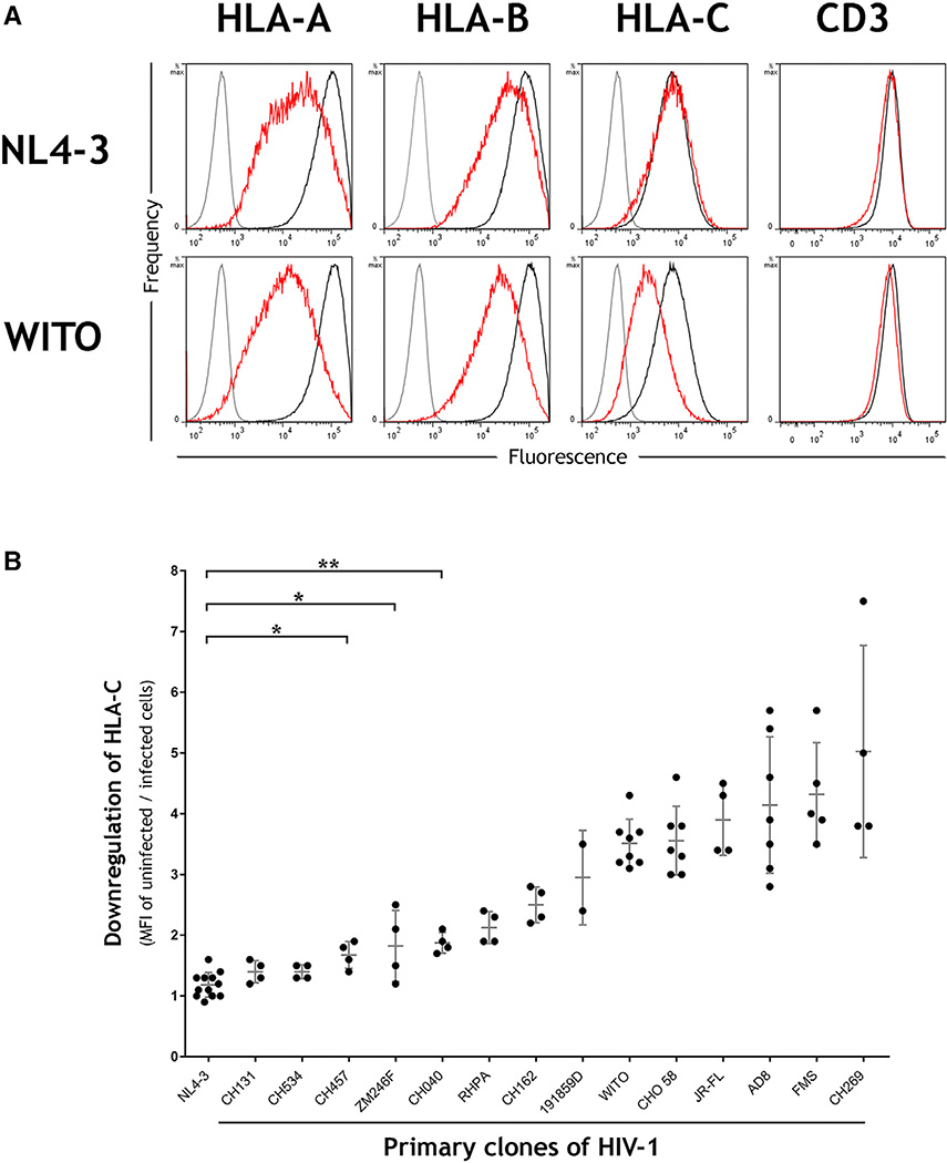
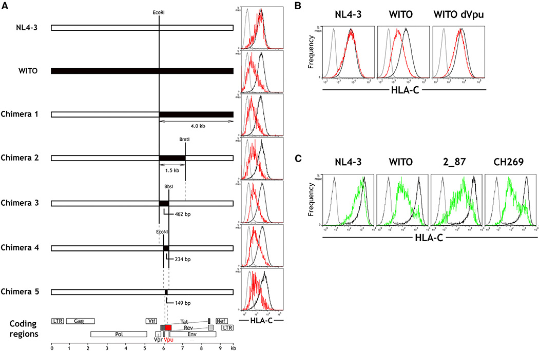
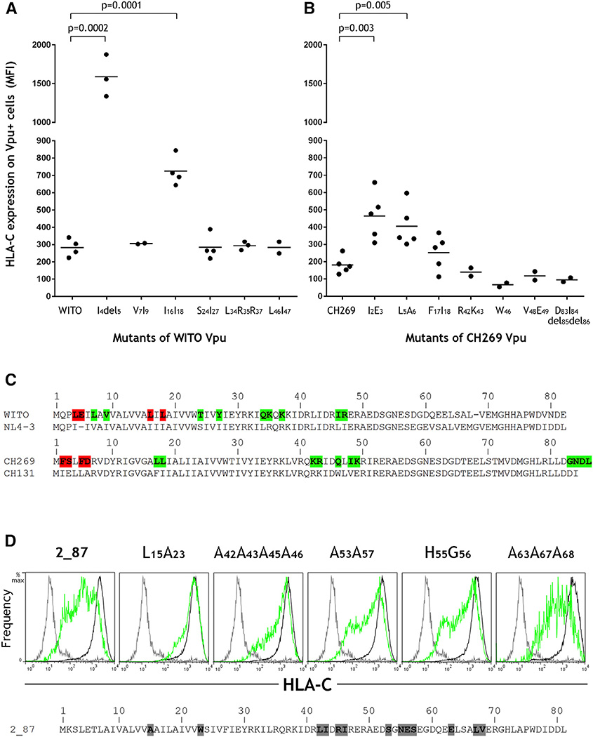
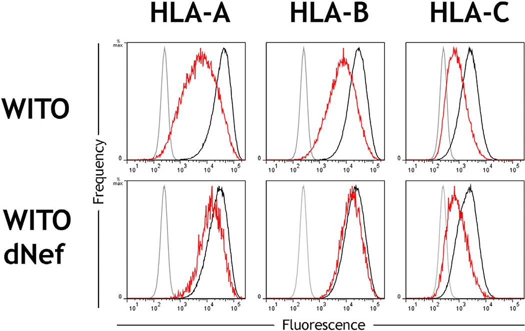
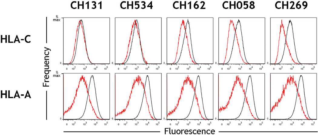
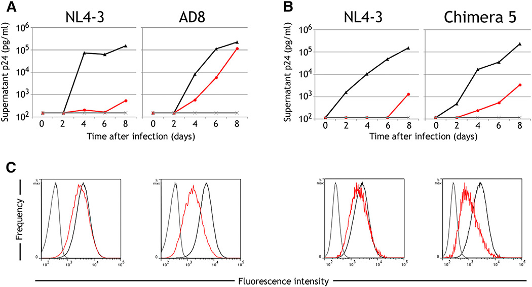
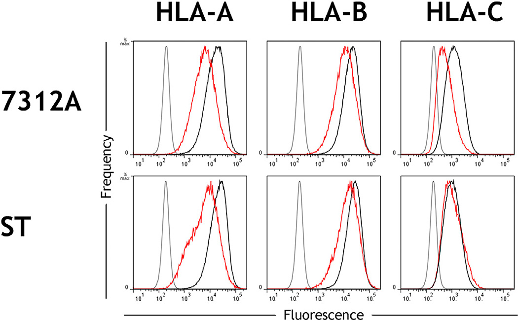
Many pathogens evade cytotoxic T lymphocytes (CTLs) by downregulating HLA molecules on infected cells, but the loss of HLA can trigger NK cell-mediated lysis. HIV-1 is thought to subvert CTLs while preserving NK cell inhibition by Nef-mediated downregulation of HLA-A and -B but not HLA-C molecules. We find that HLA-C is downregulated by most primary HIV-1 clones, including transmitted founder viruses, in contrast to the laboratory-adapted NL4-3 virus. HLA-C reduction is mediated by viral Vpu and reduces the ability of HLA-C restricted CTLs to suppress viral replication in CD4+ cells in vitro. HLA-A/B are unaffected by Vpu, and primary HIV-1 clones vary in their ability to downregulate HLA-C, possibly in response to whether CTLs or NK cells dominate immune pressure through HLA-C. HIV-2 also suppresses HLA-C expression through distinct mechanisms, underscoring the immune pressure HLA-C exerts on HIV. This viral immune evasion casts new light on the roles of CTLs and NK cells in immune responses against HIV.


Figures

Figure 1
Figure 1. HLA-C Is Downregulated by Most Primary Clones of HIV-1 in Contrast to the Laboratory-Adapted Clone NL4-3
(A) Primary CD4+ cells were infected in vitro with the HIV-1 molecular clones NL4-3 or WITO. Flow cytometry staining of HLA-A, HLA-B, HLA-C, or CD3 is shown for infected (red) or uninfected cells (black) and an isotype control (gray). Infected cells within cultures were discriminated by co-staining the viral protein Gag, and CD4 cell donors were homozygous for the A*02:01, B*44:02, C*0501 haplotype. (B) Downregulation of HLA-C is a common feature among primary clones of HIV-1. Primary CD4+ cells were infected in vitro with a panel of HIV-1 infectious molecular clones (x axis), and HLA-C downregulation was determined by flow cytometry (y axis). MFI indicates median fluorescence intensity. Plotted points represent independent infections and include donors with different HLA types. Error bars show ± 1 SD. Statistical comparisons used unpaired t tests where *p < 0.005 and **p < 0.0001, and each virus to the right of CH040 also differs from NL4-3 with p < 0.0001. See also Figures S1, S2, and S5 and Tables S1 and S2.
Figure 2
Figure 2. The HIV-1 Protein Vpu Is Responsible for Downregulation of HLA-C
(A) Full-length HIV-1 genomes were constructed comprising sequences from NL4-3 (white) and WITO (black). Flow cytometry plots show staining of isotype control (gray) and HLA-C on HIV+ (red) or HIV− (black) HeLa cells after transfection with the corresponding HIV-1 constructs and HIV+/− cell discrimination by staining viral Gag. In the final chimera 5, the 149-bp region from WITO encodes part of Vpu. (B) Primary CD4+ cells were infected in vitro with molecular clones of NL4-3, WITO, or WITO with the Vpu initiation site ablated. Flow cytometry staining for HLA-C on infected (red) and uninfected cells (black) is shown compared with an isotype control (gray), where HIV+ cells were discriminated by staining viral Gag. (C) Vpu genes cloned from NL4-3 or the primary HIV-1 subtype B viruses WITO and 2_87 and the primary subtype C virus CH269 were expressed in HeLa cells. Flow cytometry staining for HLA-C on Vpu+ (green) or Vpu− (black) cells discriminated by co-transfection with a GFP-expressing plasmid is shown compared with an isotype control (gray) and is representative of three independent experiments. See also Figures S3 and S4.
Figure 3
Figure 3. Vpu Amino Acid Residues that Affect HLA-C Downregulation
(A) Mutants of the Vpu gene cloned from the primary HIV clone WITO were expressed in HeLa cells and analyzed by flow cytometry for their ability to downregulate HLA-C as shown in Figure 2C. Mutants included each of the differences in Vpu protein sequences between WITO and NL4-3 in the region shown to discriminate HLA-C reduction as shown in Figure 2A (i.e., 5′ of the BbsI site). (B) Mutants of the Vpu gene cloned from the primary HIV clone CH269 were tested in the same way for their ability to downregulate HLA-C. Mutants included each of the differences in Vpu protein sequences between CH269 and CH131. (C) Vpu sequences from the viruses analyzed are shown, where residues implicated in HLA-C reduction are highlighted red and all others tested but showing little to no effects are highlighted green. Statistical comparisons were determined using an unpaired t test. (D) The Vpu gene cloned from the primary HIV strain 2_87 and its mutants known to affect CD4 and/or tetherin downregulation were expressed in HeLa cells. Flow cytometry staining for HLA-C on Vpu+ (green) or Vpu− (black) cells discriminated by co-transfection with a GFP-expressing plasmid is shown compared with an isotype control (gray), with data shown representative of three independent experiments. The wild-type 2_87 Vpu sequence is shown, with mutants detailed above each cytometry plot.
Figure 4
Figure 4. Vpu Downregulates HLA-C Specifically and Not HLA-B or HLA-A
Primary CD4+ cells were infected in vitro with the HIV molecular clone WITO (top) or a mutant of WITO that does not express Nef (bottom). Cytometry staining of HLA-A, HLA-B, or HLA-C is shown for infected (red) and uninfected cells (black) and for an isotype control (gray). Infected cells within cultures were discriminated by co-staining for the viral protein Gag. CD4 cells were derived from donors homozygous for the A*02:01, B*44:02, C*0501 haplotype. See also Figure S5.
Figure 5
Figure 5. Primary HIV-1 Clones Show Greater Variation in Downregulation of HLA-C Than HLA-A
Primary CD4+ cells were infected in vitro with the HIV-1 primary molecular clones CH131, CH534, CH162, CH058, and CH269. Flow cytometry staining of HLA-C (top) or HLA-A (bottom) is shown for infected cells (red) and uninfected cells (black), which were discriminated within a culture by co-staining of the viral protein Gag. Data are representative of experiments using CD4 cells from donors with multiple HLA-A and -C genotypes.
Figure 6
Figure 6. Viral Downregulation of HLA-C Impairs T Cell Inhibition of Viral Replication In Vitro
(A and B) Primary CD4+ cells expressing HLA-C* 03:04 were infected in vitro with the HIV-1 molecular clones NL4-3 and AD8 (A) or NL4-3 and chimera 5, the mutant of NL4-3 containing a 149-bp segment substituted from WITO (B). Viral replication quantified over the course of 8 days is shown for CD4 cells cultured alone (black), cocultured with an Env-specific HLA-C*03-restricted CTL clone at a CTL:CD4 ratio of 1:4 (red) and uninfected cultures (gray). Results are representative of CTL:CD4 ratios from 1:1 to 1:20 and four replicate experiments using multiple CD4 donors. (C) Flow cytometry staining of HLA-C on infected (red) or uninfected cells (black) and an isotype control (gray) is shown for each of the above experiments at day 6 post-infection, with infected cells in a culture discriminated by co-staining the viral protein Gag. See also Figure S4.
Figure 7
Figure 7. HLA-C Can Be Downregulated by Clones of HIV-2
Primary CD4+ cells were infected in vitro with the HIV-2 molecular clones 7312A or ST. Flow cytometry staining of HLA-A, HLA-B, or HLA-C is shown for infected (red) or uninfected cells (black) and an isotype control (gray). Infected cells within cultures were discriminated by CD4 downregulation among CD3+ CD8− cells. CD4 cell donors were homozygous for the A*02:01, B*44:02, C*0501 haplotype.

Comment in

  • HLA-C Downmodulation by HIV-1 Vpu.
    Barker E, Evans DT. Barker E, et al. Cell Host Microbe. 2016 May 11;19(5):570-1. doi: 10.1016/j.chom.2016.04.023. Cell Host Microbe. 2016. PMID: 27173922

References

    1. Adachi A, Gendelman HE, Koenig S, Folks T, Willey R, Rabson A, Martin MA. Production of acquired immunodeficiency syndrome-associated retrovirus in human and nonhuman cells transfected with an infectious molecular clone. J. Virol. 1986;59:284–291. - PMC - PubMed
    1. Adnan S, Balamurugan A, Trocha A, Bennett MS, Ng HL, Ali A, Brander C, Yang OO. Nef interference with HIV-1-specific CTL antiviral activity is epitope specific. Blood. 2006;108:3414–3419. - PMC - PubMed
    1. Alter G, Heckerman D, Schneidewind A, Fadda L, Kadie CM, Carlson JM, Oniangue-Ndza C, Martin M, Li B, Khakoo SI, et al. HIV-1 adaptation to NK-cell-mediated immune pressure. Nature. 2011;476:96–100. - PMC - PubMed
    1. Apps R, Murphy SP, Fernando R, Gardner L, Ahad T, Moffett A. Human leukocyte antigen (HLA) expression of primary trophoblast cells and placental cell lines, determined using single antigen beads to characterize allotype specificities of anti-HLA antibodies. Immunology. 2009;127:26–39. - PMC - PubMed
    1. Apps R, Qi Y, Carlson JM, Chen H, Gao X, Thomas R, Yuki Y, Del Prete GQ, Goulder P, Brumme ZL, et al. Influence of HLA-C expression level on HIV control. Science. 2013;340:87–91. - PMC - PubMed

MeSH terms

Substances