Review
. 2016 Jul;36(7):1316-22.
doi: 10.1161/ATVBAHA.116.306925. Epub 2016 May 12.

Factor XI as a Therapeutic Target

Affiliations
Review

Factor XI as a Therapeutic Target

David Gailani et al. Arterioscler Thromb Vasc Biol. 2016 Jul.

Abstract

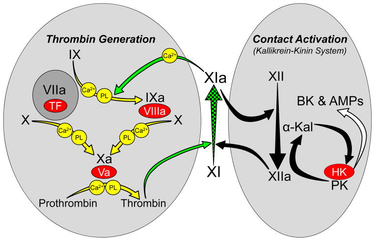
Factor XIa is a plasma serine protease that contributes to thrombin generation primarily through proteolytic activation of factor IX. Traditionally considered part of the intrinsic pathway of coagulation, several lines of evidence now suggest that factor XIa serves as an interface between the vitamin-K-dependent thrombin generation mechanism and the proinflammatory kallikrein-kinin system, allowing the 2 systems to influence each other. Work with animal models and results from epidemiological surveys of human populations support a role for factor XIa in thromboembolic disease. These data and the clinical observation that deficiency of factor XI, the zymogen of factor XIa, produces a relatively mild bleeding disorder suggest that drugs targeting factor XI or XIa could produce an antithrombotic effect while leaving hemostasis largely intact. Results of a recent trial comparing antisense-induced factor XI reduction to standard-dose low molecular-weight heparin as prophylaxis for venous thrombosis during knee replacement are encouraging in this regard. Here, we discuss recent findings on the biochemistry, physiology, and pathology of factor XI as they relate to thromboembolic disease.

Keywords: factor XI; factor XII; hemorrhage; thrombin; thrombosis.


Figures

Figure 1
Figure 1. The Relationship of Factor XI to Thrombin Generation and Contact Activation
Proteolytic reactions required for thrombin generation at an injury site are shown in the gray oval on the left, with each reaction indicated by a yellow arrow. The factor VIIa/tissue factor (TF) complex initiates thrombin generation by activating factors X and IX. Activated factor X (factor Xa) is responsible for cleaving prothrombin to form thrombin. Protease zymogens are indicated in black, and their active forms are indicated by a lower case “a”. Cofactors are shown as red ovals. Calcium (Ca2+) and phospholipid (PL) dependent reactions are indicated. Thrombin generated early in coagulation converts fXI to fXIa, which sustains thrombin production through factor IX activation (green arrows). Note that fXI activation does not require fXIIa, explaining why fXII deficiency does not cause bleeding. Proteolytic reactions involved in contact activation are shown in the gray oval on the right, with each reaction indicated by a black arrow. Artificial or abnormal surfaces facilitate fXII autoactivation. FXIIa converts prekallikrein (PK) to α-kallikrein, which activates additional fXII and cleaves high-molecular-weight kininogen (HK), liberating bradykinin (BK) and antimicrobial peptides (AMPs). Contact activation can promote thrombin generation through fXIIa-mediated activation of FXI [11,12]. There is evidence that fXIa, in turn, can activate fXII [13], although this is not a standard part of contact activation models. In plasma, PK and fXI circulate as complexes with HK, which may serve as a cofactor for PK and fXI activation. Activation of fXI by fXIIa is not required for hemostasis, but contributes to thrombosis in animal models. FXI is considered a component of contact activation (kallikrein-kinin) and thrombin generation in the scheme shown here, functioning as a bidirectional interface between the two systems. Hypothetically, activation of either system could activate the other through fXI conversion to fXIa. Image adapted from references and .
Figure 2
Figure 2. Ferric chloride-induced carotid artery occlusion
Carotid artery occlusion was induced in wild type (WT) C57Bl/6 mice, and in mice lacking factor IX (IX−/−), factor XI (fXI−/−), fXII (fXII−/−), prekallikrein (PK−/−), or high molecular weight kininogen (HK−/−) with varying concentrations of FeCl3 as indicated at the bottom of the graph. The percent of animals with patent arteries 30 minutes after FeCl3 exposure is shown (n = 10 for each bar). HK−/− mice are homozygous null for deletions of the Kng1 gene. Mice, unlike humans, have two kininogen genes (Kng1 and Kng2). Kng1 is though to be responsible for most, if not all, of the HK in plasma. Image from data in references –.
Figure 3
Figure 3. Acute vascular graft thrombosis
Thrombosis is induced by deployment of vascular grafts with collagen- or TF-coated segments into chronic arteriovenous shunts in baboons. Graft platelet content during blood perfusion and fibrin content after graft removal were measured in naïve and antibody-treated animals. Meta-analysis of experiments with antibodies that target specific reactions involving fXI and fXII indicate that inhibition of fXIa and inhibition of fXI activation by fXIIa have different effects on thrombus formation, with the most pronounced antithrombotic effect obtained with antibodies that inhibit factor IX activation by fXIa. Left Panel - values for total fibrin content and platelet deposition over the 60 minutes of each study, and maximum, average and final (last 20 minutes of the study) platelet deposition rates are shown as percentages of baseline values in the same study subject prior to antibody treatment. Red bars show mean values for animals treated with polyclonal anti-fXI antibody or the monoclonal IgG O1A6, both of which block factor IX activation by fXIa. The black bars show mean values for animals treated with monoclonal IgG 14E11 (or its humanized counterpart 3G3) which binds to fXI and specifically blocks its activation by fXIIa without interfering with fXIa activity, or with the anti-fXII monoclonal antibody 15H8, which blocks fXII activation. Error bars represent +/− one SD. Right Panel – platelet thrombus growth over time in animals treated with anti-fXI or anti-fXII antibodies (red curves) compared to untreated animals (black curves). The value for platelet deposition 60 minutes after the start of the experiment in untreated animals was arbitrarily assigned a value of 100%. Images derived from data in references and –.

References

    1. Crowther MA, Warkentin TE. Bleeding risk and the management of bleeding complications in patients undergoing anticoagulant therapy: focus on new anticoagulant agents. Blood. 2008;111:4871–4879. - PubMed
    1. Hirsh J, O’Donnell M, Weitz JI. New Anticoagulants. Blood. 2005;105:453–463. - PubMed
    1. Yeh CH, Hogg K, Weitz JI. Overview of the new oral anticoagulants: opportunities and challenges. Arterioscler Thromb Vasc Biol. 2015;35:1056–1065. - PubMed
    1. Gailani D, Bane CE, Gruber A. Factor XI and contact activation as targets for antithrombotic therapy. J Thromb Haemost. 2015;13:1383–1395. - PMC - PubMed
    1. Key NS. Epidemiologic and clinical data linking factors XI and XII to thrombosis. Hematology Am Soc Hematol Educ Program. 2014;2014:66–70. - PubMed

Publication types