Review
. 2016 Jun;14(6):360-73.
doi: 10.1038/nrmicro.2016.45. Epub 2016 May 13.

Viral evasion of intracellular DNA and RNA sensing

Affiliations
Review

Viral evasion of intracellular DNA and RNA sensing

Ying Kai Chan et al. Nat Rev Microbiol. 2016 Jun.

Abstract

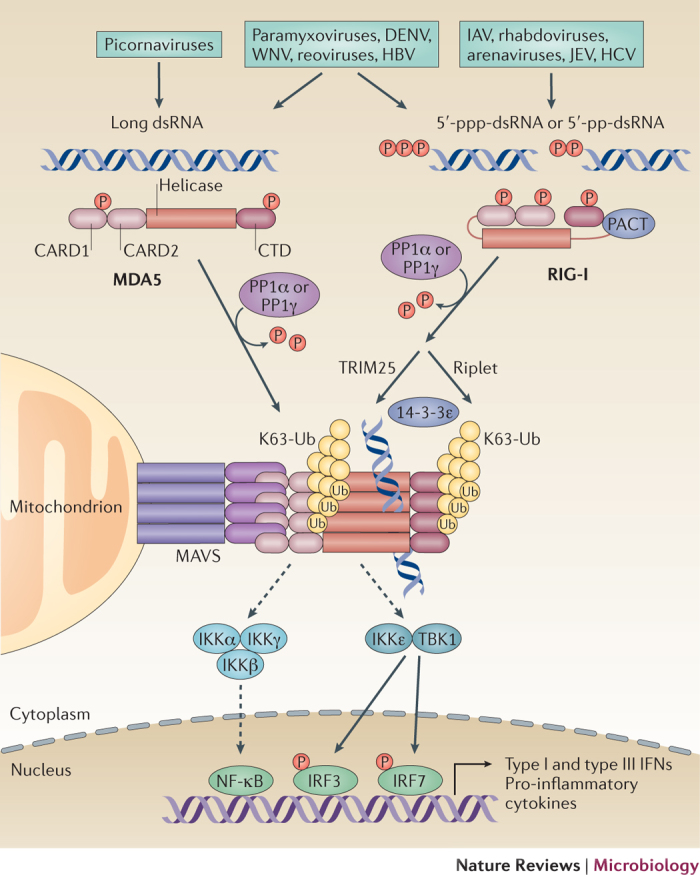
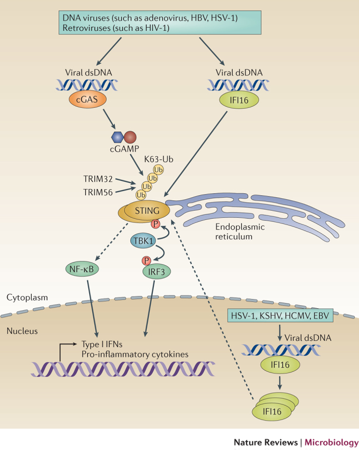
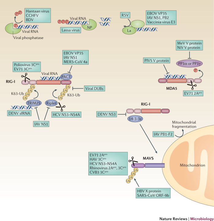
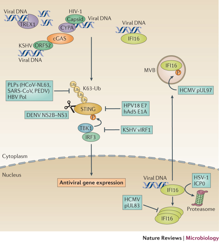
The co-evolution of viruses with their hosts has led to the emergence of viral pathogens that are adept at evading or actively suppressing host immunity. Pattern recognition receptors (PRRs) are key components of antiviral immunity that detect conserved molecular features of viral pathogens and initiate signalling that results in the expression of antiviral genes. In this Review, we discuss the strategies that viruses use to escape immune surveillance by key intracellular sensors of viral RNA or DNA, with a focus on RIG-I-like receptors (RLRs), cyclic GMP-AMP synthase (cGAS) and interferon-γ (IFNγ)-inducible protein 16 (IFI16). Such viral strategies include the sequestration or modification of viral nucleic acids, interference with specific post-translational modifications of PRRs or their adaptor proteins, the degradation or cleavage of PRRs or their adaptors, and the sequestration or relocalization of PRRs. An understanding of viral immune-evasion mechanisms at the molecular level may guide the development of vaccines and antivirals.


Conflict of interest statement

The authors declare no competing financial interests.

Figures

Figure 1
Figure 1. RLR–MAVS-mediated signal transduction pathway.
RIG-I-like receptors (RLRs) have been identified as important cytoplasmic viral RNA sensors that recognize the genomic RNA and/or RNA replication intermediates of numerous viruses. The two RLR members retinoic acid-inducible gene-I protein (RIG-I) and melanoma differentiation-associated protein 5 (MDA5) are kept inactive in uninfected cells through the phosphorylation of their caspase activation and recruitment domains (CARDs) and carboxy-terminal domains (CTDs). In addition, RIG-I adopts a 'closed' auto-inhibited conformation. Following viral infection, RIG-I recognizes cytoplasmic viral short double-stranded RNA (dsRNA) that contains a 5′-triphosphate or 5′-diphosphate moiety, whereas MDA5 detects long dsRNA structures. Following the binding of RNA, RIG-I and MDA5 are dephosphorylated by PP1α or PP1γ, which induces the formation of a signalling-active CARD conformation. RIG-I is further activated by the Lys63-linked ubiquitylation of its CARDs that is mediated by tripartite motif protein 25 (TRIM25). Riplet, another E3 ubiquitin ligase, mediates the Lys63-linked ubiquitylation of the CTD of RIG-I, which is also crucial for the activation of RIG-I. Lys63-linked ubiquitylation induces the tetramerization of RIG-I (the signalling-active form of RIG-I), which subsequently interacts with the adaptor mitochondrial antiviral signalling protein (MAVS) on mitochondria, mitochondria-associated membranes (MAMs) or peroxisomes (not shown). The mitochondrial-targeting chaperone protein 14-3-3ɛ is essential for the translocation of RIG-I to mitochondrial MAVS. In the case of MDA5, binding to long dsRNA induces MDA5 filament formation, which subsequently enables MDA5 to bind to MAVS. MAVS activates TBK1 or IκB kinase-ɛ (IKKɛ) as well as the IKKα–IKKβ–IKKγ complex, which activate interferon (IFN) regulatory factor 3 (IRF3) and IRF7, and nuclear factor-κB (NF-κB), respectively, through phosphorylation events. IRF3 and/or IRF7 and NF-κB together with activator protein 1 (AP1; not illustrated) induce the gene expression of type I IFNs (mainly IFNα subtypes and IFNβ), type III IFNs (IFNλ subtypes), and many other pro-inflammatory cytokines and chemokines, to establish an antiviral state. Solid arrows indicate direct signalling events. Dashed arrows indicate indirect signalling events. DENV, dengue virus; HBV, hepatitis B virus; HCV, hepatitis C virus; IAV, influenza A virus; JEV, Japanese encephalitis virus; K63-Ub, Lys63-linked ubiquitylation; P, phosphate; PACT, protein kinase R activator; Ub, ubiquitin; WNV, West Nile virus. PowerPoint slide
Figure 2
Figure 2. Antiviral signalling mediated by cGAS and IFI16 through STING.
In the cytoplasm of infected cells, cyclic GMP–AMP synthase (cGAS) recognizes double-stranded DNA (dsDNA) from DNA viruses or dsDNA that is produced by retroviruses through the reverse transcription of their RNA genomes. Following the binding of DNA, cGAS synthesizes the second messenger cyclic GMP–AMP (cGAMP), which then binds to and activates stimulator of interferon (IFN) genes (STING) on the endoplasmic reticulum. STING is further activated by dimerization and Lys63-linked ubiquitylation that is mediated by tripartite motif protein 32 (TRIM32) and TRIM56. Furthermore, STING is phosphorylated by TBK1. The sensor IFNγ-inducible protein 16 (IFI16) senses viral dsDNA in both the cytoplasm and the nucleus. Following the binding of viral DNA, IFI16 multimerizes and then signals through STING in the cytoplasm. The activation of STING induces the expression of type I IFN genes and other pro-inflammatory cytokines through the TBK1–IFN regulatory factor 3 (IRF3) axis and nuclear factor-κB (NF-κB). Solid arrows indicate well-established signalling events. Dashed arrows indicate signalling events that are indirect or that have not yet been fully elucidated. EBV, Epstein–Barr virus; HBV, hepatitis B virus; HCMV, human cytomegalovirus; HSV-1, herpes simplex virus 1; K63-Ub, Lys63-linked ubiquitylation; KSHV, Kaposi sarcoma-associated herpesvirus; P, phosphate; Ub, ubiquitin. PowerPoint slide
Figure 3
Figure 3. Viral immune evasion of RLR–MAVS signalling.
Most successful viral pathogens are equipped with effective strategies to evade or inhibit the activation of intracellular pattern recognition receptors (PRRs), such as retinoic acid-inducible gene-I protein (RIG-I) or melanoma differentiation-associated protein 5 (MDA5), or the activation of their adaptor mitochondrial antiviral signalling protein (MAVS). To prevent the activation of RIG-I, viral phosphatases can process the 5′-triphosphate moiety in the viral RNA, or viral nucleases, such as the nucleoprotein (NP) of Lassa virus, can digest free double-stranded RNA (dsRNA). Furthermore, viral proteins, such as viral protein 35 (VP35) from EBOV, non-structural protein 1 (NS1) or PB2 from influenza A virus (IAV) and the E3 protein from vaccinia virus, or host proteins (such as La) bind to viral RNA to inhibit the recognition of pathogen-associated molecular patterns (PAMPs) by RIG-I. Several viruses manipulate specific post-translational modifications of RIG-I and/or MDA5, thereby blocking their signalling abilities. For example, viruses prevent the Lys63-linked ubiquitylation of RIG-I by encoding viral deubiquitylating enzymes (DUBs). NS1 from IAV and the NS3–NS4A protease complex from hepatitis C virus (HCV) antagonize the cellular E3 ubiquitin ligases, tripartite motif protein 25 (TRIM25) and/or Riplet, thereby also inhibiting RIG-I ubiquitylation and thus its activation. Furthermore, subgenomic flavivirus RNA (sfRNA) from dengue virus (DENV) binds to TRIM25 to block sustained RIG-I signalling. To suppress the activation of MDA5, the V proteins from measles virus (MeV) and Nipah virus (NiV) prevent the PP1α-mediated or PP1γ-mediated dephosphorylation of MDA5, keeping it in its phosphorylated inactivate state, whereas the V protein of parainfluenza virus 5 (PIV5) blocks the ATPase activity of MDA5. Furthermore, VP35 from EBOV, NS1 from IAV and the 4a protein from Middle East respiratory syndrome coronavirus (MERS-CoV) target protein kinase R activator (PACT) to antagonize RIG-I. The NS3 protein from DENV targets the trafficking factor 14-3-3ɛ to prevent the translocation of RIG-I to MAVS at the mitochondria. Numerous viruses encode proteases (Pro) to cleave RIG-I, MDA5 and/or MAVS. PB1-F2 from IAV translocates into the mitochondrial inner membrane space to accelerate mitochondrial fragmentation. Other viruses subvert cellular degradation pathways to inhibit RLR–MAVS-dependent signalling. Specifically, the X protein from hepatitis B virus (HBV) and the 9b protein from severe acute respiratory syndrome (SARS)-associated coronavirus (SARS-CoV) promote the ubiquitylation and degradation of MAVS. BDV, Borna disease virus; CCHFV, Crimean–Congo haemorrhagic fever virus; CVB3, coxsackievirus B3; EV71, enterovirus 71; HAV, hepatitis A virus; K63-Ub, Lys63-linked ubiquitylation; P, phosphate; RSV, respiratory syncytial virus; Ub, ubiquitin. PowerPoint slide
Figure 4
Figure 4. Viral immune evasion of cGAS, IFI16 and STING.
DNA viruses have developed molecular strategies to evade or inhibit intracellular DNA sensors and escape immune signalling during both acute and persistent infection. To prevent the activation of cyclic GMP–AMP synthase (cGAS), HIV-1 uses the cellular 3′-repair exonuclease 1 (TREX1) to degrade excess reverse transcribed viral DNA. In addition, the HIV-1 capsid recruits host-encoded factors, such as cyclophilin A (CYPA), which prevent the sensing of reverse transcribed DNA by cGAS. Furthermore, the tegument protein ORF52 of Kaposi sarcoma-associated herpesvirus (KSHV) binds to both viral DNA and cGAS to inhibit the activity of cGAS. To antagonize the activation of stimulator of interferon (IFN) genes (STING), the polymerase (Pol) of hepatitis B virus (HBV) and the papain-like proteases (PLPs) of human coronavirus NL63 (HCoV-NL63), severe acute respiratory syndrome (SARS)-associated coronavirus (SARS-CoV) and porcine epidemic diarrhoea virus (PEDV), prevent or remove the Lys63-linked ubiquitylation of STING. The viral IFN regulatory factor 1 (vIRF1) protein of KSHV blocks the TBK1-mediated phosphorylation of STING and thereby its signalling ability. The E7 protein of human papillomavirus 18 (HPV18) and the E1A protein of human adenovirus 5 (hAd5) bind to STING and inhibit its activation, whereas the NS2B–NS3 protease of dengue virus (DENV) cleaves STING to inactivate it. To inhibit IFNγ-inducible protein 16 (IFI16), infected cell protein 0 (ICP0) of herpes simplex virus 1 (HSV-1) targets IFI16 in the nucleus for proteasomal degradation, whereas the tegument protein pUL83 of human cytomegalovirus (HCMV) binds to IFI16 to prevent its oligomerization and thus its activation. The pUL97 protein of HCMV phosphorylates IFI16 during viral replication and relocalizes it from the nucleus to multivesicular bodies (MVBs). K63-Ub, Lys63-linked ubiquitylation; P, phosphate; Ub, ubiquitin. PowerPoint slide

Similar articles

Cited by

References

    1. Takeuchi O, Akira S. Pattern recognition receptors and inflammation. Cell. 2010;140:805–820. doi: 10.1016/j.cell.2010.01.022. - DOI - PubMed
    1. Dambuza IM, Brown GD. C-type lectins in immunity: recent developments. Curr. Opin. Immunol. 2015;32:21–27. - PMC - PubMed
    1. Goubau D, Deddouche S, Reis e Sousa C. Cytosolic sensing of viruses. Immunity. 2013;38:855–869. - PMC - PubMed
    1. Schneider WM, Chevillotte MD, Rice CM. Interferon-stimulated genes: a complex web of host defenses. Annu. Rev. Immunol. 2014;32:513–545. - PMC - PubMed
    1. Yoneyama M, et al. The RNA helicase RIG-I has an essential function in double-stranded RNA-induced innate antiviral responses. Nat. Immunol. 2004;5:730–737. - PubMed

MeSH terms