. 2016 Jul 1;415(1):75-86.
doi: 10.1016/j.ydbio.2016.05.001. Epub 2016 May 10.

Drosophila male and female germline stem cell niches require the nuclear lamina protein Otefin

Affiliations

Drosophila male and female germline stem cell niches require the nuclear lamina protein Otefin

Lacy J Barton et al. Dev Biol. .

Abstract

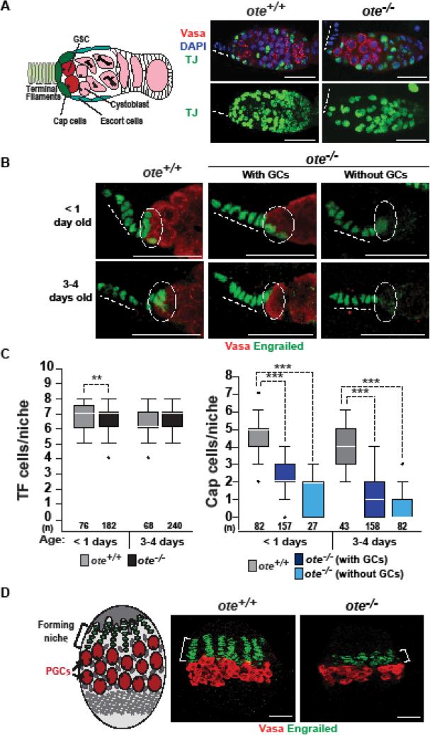
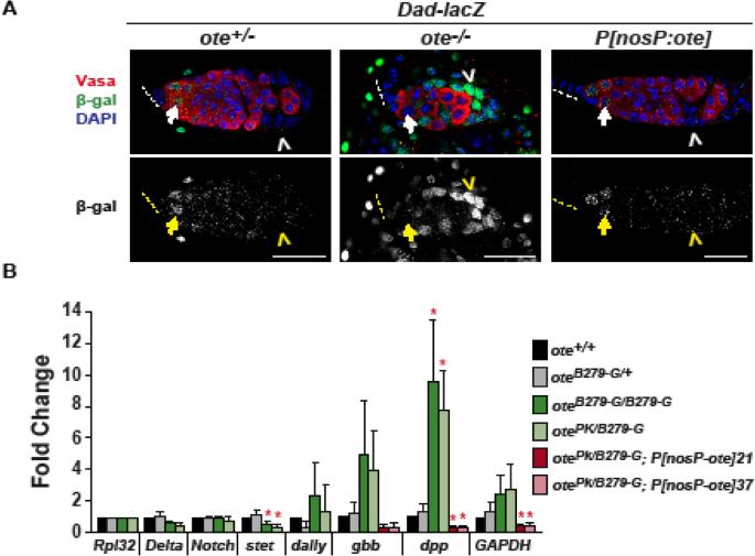
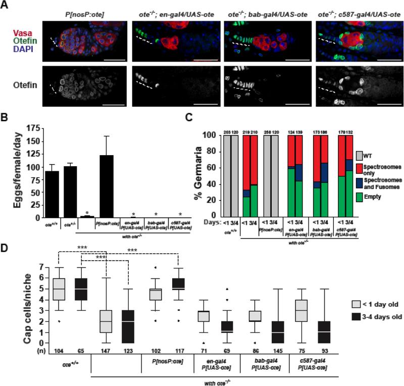
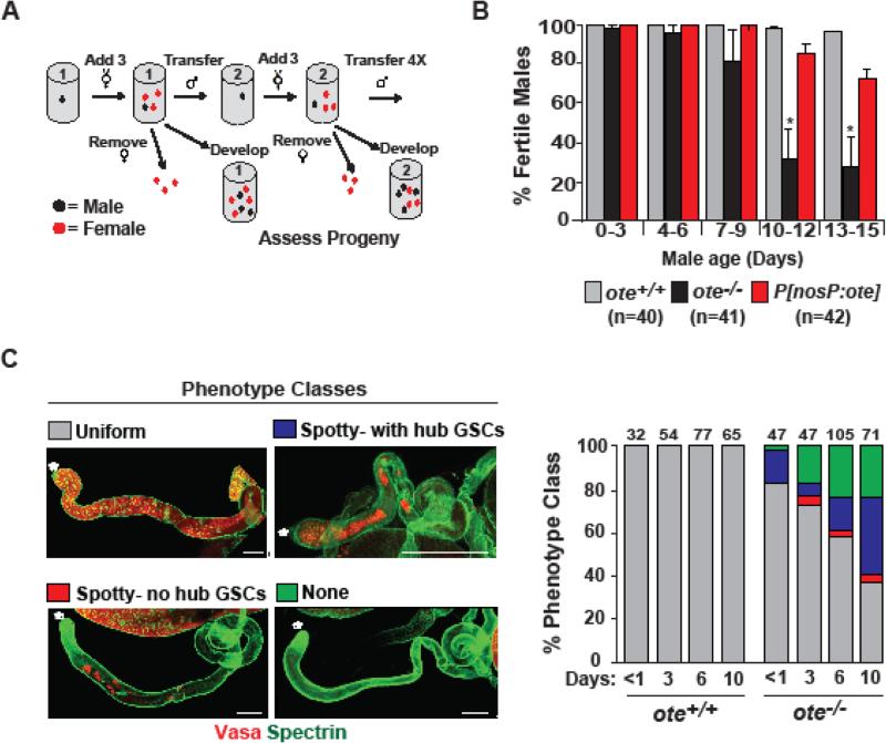
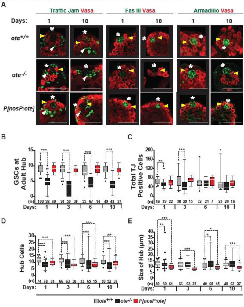
The nuclear lamina is an extensive protein network that underlies the inner nuclear envelope. This network includes the LAP2-emerin-MAN1-domain (LEM-D) protein family, proteins that share an association with the chromatin binding protein Barrier-to-autointegration factor (BAF). Loss of individual LEM-D proteins causes progressive, tissue-restricted diseases, known as laminopathies. Mechanisms associated with laminopathies are not yet understood. Here we present our studies of one of the Drosophila nuclear lamina LEM-D proteins, Otefin (Ote), a homologue of emerin. Previous studies have shown that Ote is autonomously required for the survival of female germline stem cells (GSCs). We demonstrate that Ote is also required for survival of somatic cells in the ovarian niche, with loss of Ote causing a decrease in cap cell number and altered signal transduction. We show germ cell-restricted expression of Ote rescues these defects, revealing a non-autonomous function for Ote in niche maintenance and emphasizing that GSCs contribute to the maintenance of their own niches. Further, we investigate the requirement of Ote in the male fertility. We show that ote mutant males become prematurely sterile as they age. Parallel to observations in females, this sterility is associated with GSC loss and changes in somatic cells of the niche, phenotypes that are largely rescued by germ cell-restricted Ote expression. Taken together, our studies demonstrate that Ote is required autonomously for survival of two stem cell populations, as well as non-autonomously for maintenance of two somatic niches. Finally, our data add to growing evidence that LEM-D proteins have critical roles in stem cell survival and tissue homeostasis.

Keywords: Drosophila; LEM-domain proteins; Nuclear lamina; Oogenesis, Germline stem cells; Spermatogenesis.


Figures

Figure 1
Figure 1. Loss of Ote disrupts somatic cells in the germarium
A. Left: Schematic of the ovarian stem cell niche, showing somatic cells that include terminal filament (TF) cells (light green), cap cells (dark green), escort cells (blue), and germ cells that include germline stem cells (GSCs; red), cytoblasts and differentiating germ cells (pink). Right: Confocal images of ote+/+ and ote−/− germaria stained with antibodies against TJ (detects cap and escort cells, green), Vasa (detects germ cells, red), and DNA stained with DAPI (blue). Panels on the top are single confocal slices of a Z-stack through an entire germarium. Images on the bottom are maximum projections of each slice in the Z-stack, showing all TJ-positive cells in the germarium. Dashed lines indicate position of the TF. Scale bars, 25 μm. B. Confocal images of one- and three-to-four-day-old ote+/+ and ote−/− germaria stained with antibodies against Vasa and Engrailed (detects TF and cap cells). Dotted circles indicate the position of cap cells. C. Quantification of the number of TF cells and cap cells present within ote+/+ (gray) and ote−/− germaria with (black-C or dark blue-D) or without GSCs (light blue). The number of niches analyzed is shown below each bar. For each box, the box represents the 25th to 75th percentile interval, the line represents the median, and the whiskers represent the 5th to 95th percentile interval and non-outlier range. The Mann-Whitney-Wilcoxon Test was used to determine statistics (*p<0.05; **p<0.01; and ***p<0.001). D. Left: Schematic of the developing female larval gonad, showing TF cells (green), primordial germline stem cells (PGCs, red), and Intermingled cells (blue). The bracket highlights the region of the forming niche. Right: Confocal images ote+/+ and ote−/− stem cell niche in third instar larvae, using antibodies against Engrailed (green) and Vasa (red). Scale bars, 25 μm.
Figure 2
Figure 2. Analysis of BMP signaling in the ote−/− germaria
A. Analysis of BMP signaling in ote+/+ and ote−/− germaria using the Dad-LacZ reporter gene. This reporter carries a P element insertion of lacZ in the Daughters-against-Dpp gene (Tsuneizumi et al., 1997). Top panels show confocal images from germaria obtained from one-day-old ote+/+ and ote−/− Dad-lacZ ovaries stained for Vasa (red), β-galactosidase (green) and the DNA dye DAPI (blue). Bottom panels show confocal images of β-galactosidase staining shown in white. Arrows indicate germ cells and chevrons indicate somatic cells. Dashed lines indicate the position of the TF. Scale bars, 25 μm. B. Quantitative reverse transcription PCR of RNAs isolated from ote+/+ and ote−/− ovaries obtained from less than two-hour-old females. RNAs were obtained from females representing two ote+/+ (black, gray bars), two ote−/− (green bars) genotypes and two ote−/−, P[nosP:ote, w+] lines (red bars). RNA levels were normalized to Rpl32 and fold change was set relative to the ote+/+ genotype. Error bars indicate standard deviation from three biological replicates (* = p<0.05, Student's t-test).
Figure 3
Figure 3. Germ cell-restricted Ote expression rescues somatic niche defects in ovaries
A. Confocal images of three-day-old germaria in ovaries isolated from ote−/−; P[nosP:ote, w+] females and several one-day-old ote−/− females carrying three different cell-type restricted GAL4 drivers and the P[UASt-ote] responder. Ovaries were stained with Vasa (red), Ote (green), and DAPI (blue) in the top panels and Ote only (white) in the bottom panels. Scale bars represent 25 μm. B. Quantification of fecundity of three-day-old females of the following genotypes: ote+/+, ote+/−, ote−/−, P[nosP:ote, w+] and the indicated GAL4-P[UASt-ote] responders. Bars indicate the variation from three independent experiments. C. Quantification of germarial class prevalence in the genetic backgrounds listed in B. The total number of germaria analyzed is shown above each bar. D. Quantification of numbers of cap cells in less than one-day-old (gray) and in three-to-four-day-old (black) females of the indicated genotypes. The number of niches analyzed is shown below each bar. The limits of box plots and analyses are as described in Fig. 1.
Figure 4
Figure 4. Loss of Ote causes age-dependent male sterility
A. Schematic of sperm exhaustion assay to test fertility of aging ote−/− males. Single males (black dot) were mated to three to four females (red dots). After three days, each male was transferred to a new vial and mated to new virgin females; a process continued four times. B. Quantification of the fertility of ote+/+, ote−/−, and ote−/−, P[nosP:ote, w+] males. The number of males of each genotype is shown. Bars indicate the variation from six biological replicates (P>0.05; Student's t-test). C. Shown are confocal images of the four representative phenotypic testes classes identified using staining with Vasa and Spectrin antibodies. Testis images are oriented with anterior to the left. Asterisks mark the location of the hub. Scale bars, 200 μm. Graphs indicate the quantification of the prevalence of each phenotypic class in testes obtained from ote+/+ and ote−/− males of the indicated age.
Figure 5
Figure 5. Age-dependent changes in the adult ote−/− testis niche
A. Confocal images of one- and ten-day-old ote+/+, ote −/−, and ote−/−, P[nosP:ote, w+] testes stained with Vasa (red) and one of the following in green: TJ, Fascilin (Fas) III or Armadillo. Yellow arrowheads mark positions of GSCs; white arrowheads mark positions of CySCs; asterisks mark positions of the hubs. Scale bars, 20 μm. B-E. Quantification of the number of GSCs (B), the total number of TJ-positive cells (C), the total number of hub cells (D) and hub size (E) in aged testes obtained from ote+/+ (gray), ote−/− (black), and ote−/−, P[nosP:ote, w+] (red) males. The number of testes analyzed is shown below each bar. The limits of box plots and analyses are as described in Fig. 1.
Figure 6
Figure 6. Larval ote−/− testes show defects
A. Confocal images of ote+/+ and ote−/− third instar larval gonads. Gonads were stained with antibodies against Vasa (red), TJ (green) and costained with DAPI (blue). Yellow arrowheads mark positions of GSCs; white arrowheads mark positions of CySCs; asterisks mark positions of the hub. Scale bars, 50 μm. B-C. Quantification of the length (B) and number of GSCs (C) in ote+/+ (gray) and ote−/− (black) third instar larval gonads. The number of gonads analyzed is shown below each bar. The limits of box plots and analyses are as described in Fig. 1.

References

    1. Barkan R, Zahand AJ, Sharabi K, Lamm AT, Feinstein N, Haithcock E, Wilson KL, Liu J, Gruenbaum Y. Ce-emerin and LEM-2: essential roles in Caenorhabditis elegans development, muscle function, and mitosis. Mol Biol Cell. 2012;23:543–552. - PMC - PubMed
    1. Barton LJ, Pinto BS, Wallrath LL, Geyer PK. The Drosophila nuclear lamina protein otefin is required for germline stem cell survival. Dev Cell. 2013;25:645–654. - PMC - PubMed
    1. Barton LJ, Soshnev AA, Geyer PK. Networking in the nucleus: a spotlight on LEM-domain proteins. Curr Opin Cell Biol. 2015;34:1–8. - PMC - PubMed
    1. Barton LJ, Wilmington SR, Martin MJ, Skopec HM, Lovander KE, Pinto BS, Geyer PK. Unique and Shared Functions of Nuclear Lamina LEM Domain Proteins in Drosophila. Genetics. 2014;197:653–665. - PMC - PubMed
    1. Baxley RM, Soshnev AA, Koryakov DE, Zhimulev IF, Geyer PK. The role of the Suppressor of Hairy-wing insulator protein in Drosophila oogenesis. Dev Biol. 2011;356:398–410. - PMC - PubMed

MeSH terms