. 2016 May 12:6:25834.
doi: 10.1038/srep25834.

Homozygous mutation of VPS16 gene is responsible for an autosomal recessive adolescent-onset primary dystonia

Affiliations

Homozygous mutation of VPS16 gene is responsible for an autosomal recessive adolescent-onset primary dystonia

Xiaodong Cai et al. Sci Rep. .

Abstract

Dystonia is a neurological movement disorder that is clinically and genetically heterogeneous. Herein, we report the identification a novel homozygous missense mutation, c.156 C > A in VPS16, co-segregating with disease status in a Chinese consanguineous family with adolescent-onset primary dystonia by whole exome sequencing and homozygosity mapping. To assess the biological role of c.156 C > A homozygous mutation of VPS16, we generated mice with targeted mutation site of Vps16 through CRISPR-Cas9 genome-editing approach. Vps16 c.156 C > A homozygous mutant mice exhibited significantly impaired motor function, suggesting that VPS16 is a new causative gene for adolescent-onset primary dystonia.


Figures

Figure 1
Figure 1. Clinical characteristics of the five patients with dystonia.
Pedigree of the five patients with dystonia. Each generation is denoted by a Roman numeral, and each individual by an Arabic numeral. Empty symbols represent unaffected individuals. Solid symbols represent dystonia patients. Slashes represent deceased individuals. Double horizontal lines represent consanguious couples. The proband is indicated by arrow. (b) MRI image of the proband IV6.
Figure 2
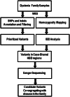
Figure 2. Flowchart of disease-causing gene exploration of dystonia patients from WES.
Figure 3
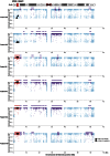
Figure 3. Distribution of identified homozygous region on chromosome 20.
Upper panel illustrates the localizations of VPS16 and SIGLEC1 on chromosome 20 (red bars). Bottom panel represents the homozygous regions on chromosome 20 identified from indicated family members. Each dot represents each SNP markers identified from WES. Purple color represents SNPs within a homozygous region, whereas red represents in a case-shared and control-absent HBD. X axis indicates the genomic location of SNP markers, whereas Y axis indicates the ratio of the number of dominant reads to the total number of reads per SNP position. Triangles and squares represents novel variants (c.156 C > A; c.1372 C > T) identified in VPS16 and SIGLEC1 respectively.
Figure 4
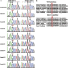
Figure 4. Sanger sequencing and protein conservation analysis of VPS16 and SIGLEC1 variants.
(a) Sequencing chromatograms are shown. Affected codons are framed in black, and variants found in individuals are indicated by arrows. (b) Protein alignment of VPS16 and SIGLEC1 variants from seven different vertebrate species using ClustalW. Altered Amino acid are indicated on the top panel, whereas Refseq residues are framed in red and indicated by arrows.
Figure 5
Figure 5. Targeted mutation of mouse Vps16 gene.
(a) Schematic representation of the genome localization of c.156 C > A mutation in Vps16 gene and construction of the targeting sgRNA and oligonucleotide. UTR regions are colored in yellow, β-Propeller domain in purple and α-Solenoid domain in blue. Mutation site is indicated by red color and black arrows. PAM motif is indicated in green. (b) Sequencing chromatograms of Vps16 c.156 C > A mutant mice. Mutation site is indicated by arrows. (c) Western blot analysis of VPS16 expression from retro-orbital blood of WT and Vps16-mutant mice. (d) Rotating rod analysis of motor function in WT and Vps16-mutant mice. Graph represents the mean ± SE obtained from at least three independent experiments. The unpaired Student’s T-test revealed a significant difference between WT (n = 5) and Vps16-mutant mice (n = 3). ***Represents p < 0.0001 compared to WT mice.

References

    1. Fahn S. Concept and classification of dystonia. Adv Neurol 50, 1–8 (1988). - PubMed
    1. Lohmann K. & Klein C. Genetics of dystonia: what’s known? What’s new? What’s next? Mov Disord 28, 899–905 (2013). - PubMed
    1. Ozelius L. J. et al. The early-onset torsion dystonia gene (DYT1) encodes an ATP-binding protein. Nat Genet 17, 40–8 (1997). - PubMed
    1. Nolte D., Niemann S. & Muller U. Specific sequence changes in multiple transcript system DYT3 are associated with X-linked dystonia parkinsonism. Proc Natl Acad Sci USA 100, 10347–52 (2003). - PMC - PubMed
    1. Hersheson J. et al. Mutations in the autoregulatory domain of beta-tubulin 4a cause hereditary dystonia. Ann Neurol 73, 546–53 (2013). - PMC - PubMed

Publication types

MeSH terms

Substances