. 2016 Jun 7;7(23):35353-68.
doi: 10.18632/oncotarget.9286.

Monomeric gremlin is a novel vascular endothelial growth factor receptor-2 antagonist

Affiliations

Monomeric gremlin is a novel vascular endothelial growth factor receptor-2 antagonist

Elisabetta Grillo et al. Oncotarget. .

Abstract

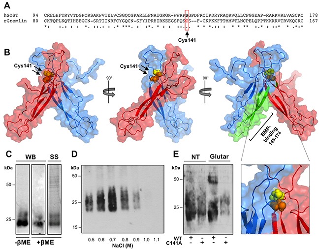
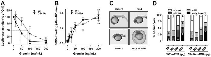
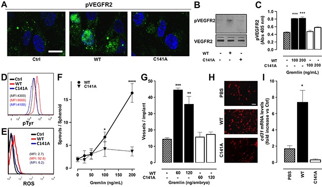
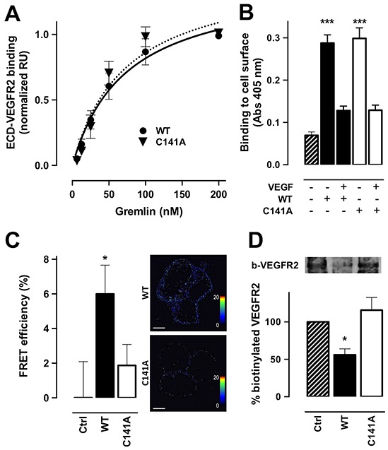
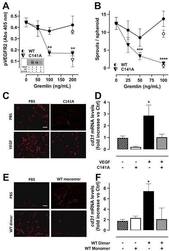
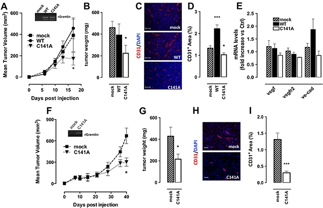
Angiogenesis plays a key role in various physiological and pathological conditions, including inflammation and tumor growth. The bone morphogenetic protein (BMP) antagonist gremlin has been identified as a novel pro-angiogenic factor. Gremlin promotes neovascular responses via a BMP-independent activation of the vascular endothelial growth factor (VEGF) receptor-2 (VEGFR2). BMP antagonists may act as covalent or non-covalent homodimers or in a monomeric form, while VEGFRs ligands are usually dimeric. However, the oligomeric state of gremlin and its role in modulating the biological activity of the protein remain to be elucidated.Here we show that gremlin is expressed in vitro and in vivo both as a monomer and as a covalently linked homodimer. Mutagenesis of amino acid residue Cys141 prevents gremlin dimerization leading to the formation of gremlinC141A monomers. GremlinC141A monomer retains a BMP antagonist activity similar to the wild-type dimer, but is devoid of a significant angiogenic capacity. Notably, we found that gremlinC141A mutant engages VEGFR2 in a non-productive manner, thus acting as receptor antagonist. Accordingly, both gremlinC141A and wild-type monomers inhibit angiogenesis driven by dimeric gremlin or VEGF-A165. Moreover, by acting as a VEGFR2 antagonist, gremlinC141A inhibits the angiogenic and tumorigenic potential of murine breast and prostate cancer cells in vivo.In conclusion, our data show that gremlin exists in multiple forms endowed with specific bioactivities and provide new insights into the molecular bases of gremlin dimerization. Furthermore, we propose gremlin monomer as a new inhibitor of VEGFR2 signalling during tumor growth.

Keywords: BMP; VEGFR2 antagonist; angiogenesis; gremlin; oligomerization.


Conflict of interest statement

The authors declare there are no relevant conflicts of interest to disclose.

Figures

Figure 1
Figure 1. Gremlin exists both as a monomer and a covalent dimer
A. total lysates from healthy murine organs were immunoprecipitated with anti-gremlin antibody, separated by SDS-PAGE under non-reducing conditions and probed with anti-gremlin antibody. Black arrow, gremlin dimer; open arrow, gremlin monomer. IgG were used as a control. B. FGF2-T-MAE cells were treated with increasing concentrations of H2O2 for 1 hour. At the end of incubation, the cells were incubated for 4 hours with fresh medium. Conditioned medium was collected and immunoprecipitated with anti-gremlin antibody. Immunoprecipitated fractions were analysed by WB under non-reducing conditions. C-D. recombinant his-tagged gremlinWT was transiently expressed in HEK293T cells, purified by IMAC and analyzed by SDS-PAGE followed by Western blotting (WB) under non-reducing (C) or by analytical size exclusion chromatography. The elution profile of gremlin was obtained by dot blot analysis of the eluted fractions (D). Black arrows indicate the retention volume of standard proteins (seroalbumin: 132 and 66 kDa; ovalbumin: 45 kDa; carbonic anhydrase: 29 kDa and lactoalbumin: 14.2 kDa). E. IMAC purified gremlin was further subjected to heparin-affinity chromatography. Heparin column was washed with a discontinuous NaCl gradient. Eluted fractions were separated by SDS-PAGE under non-reducing conditions and probed with anti-gremlin antibody. Black arrow, gremlin dimer; open arrow, gremlin monomer. F. pure dimeric gremlinWT was analysed by SDS-PAGE followed by Western blotting (WB) under non-reducing (−βME) and reducing (+βME) conditions and by silver staining (SS) of the gel.
Figure 2
Figure 2. Gremlin forms disulfide-bound homodimers through Cys141
A. CLUSTAL W sequence alignment of the cystine-knot domain of gremlin and of structurally related SOST. B. surface view of the predicted structure of gremlin homodimer generated by docking simulation with RosettaDock software (model 0267; total score: −29.594; interface score −4.429; interchain contact −20). Gremlin homodimer subunits are in red or light blue. Cys141 residues are in yellow and orange spheres. The previously identified BMP4-binding region of gremlin is depicted in green. C. gremlinC141A mutant was transiently expressed in HEK293T cells and sequentially purified from cell supernatant by IMAC and heparin-affinity chromatography. Heparin column was eluted with a discontinuous NaCl gradient. Eluted fractions were separated by SDS-PAGE under non-reducing conditions and probed with anti-gremlin antibody. D. purified gremlinC141A monomer was analysed by WB under non-reducing and reducing conditions and by SS of the gel. E. Gremlin wt and gremlinC141A were incubated in the absence or in the presence of 0.05% glutaraldehyde for 20 minutes at RT, separated by SDS-PAGE under reducing conditions and probed with anti-gremlin antibody.
Figure 3
Figure 3. GremlinC141A retains its BMP-antagonist activity
A. HepG2 cells were transiently transfected with a construct harbouring BMP-responsive hepcidin promoter upstream to the luciferase reporter gene. Serum-starved transfected cells were then stimulated with 50 ng/mL of BMP4 in the absence or the presence of increasing amounts of either gremlinWT (•) or gremlinC141A (▼). After 16 hours incubation, cells were lysed, and luciferase activity was measured. Data are expressed as percent of luciferase activity measured in BMP4 stimulated cells and are the mean ± SEM of 3 independent experiments. Two-Way ANOVA followed by Bonferroni's test revealed that the dose-response curves of the two proteins were not statistically different. B. 96-well plates coated with 250 ng/mL of BMP4 were incubated with increasing concentrations of gremlinWT or gremlinC141A. Then, the capacity of gremlinWT (•) or gremlinC141A (▼) to bind to immobilized BMP4 was assessed by ELISA assay using an anti-gremlin antibody. C-D. zebrafish embryos were injected (at 1-4 cell stage) with the indicated doses of either gremlinWT or gremlinC141A mRNA. Representative dorsalized embryo phenotypes at 24 hours after injection (C). All embryos were viewed laterally. D, percentage of embryos in different categories at 24 hours after injection. Data are the mean ± SEM of 3 independent experiments. *, p<0.05; **, p<0.01, One-Way ANOVA followed by Bonferroni's test versus control.
Figure 4
Figure 4. Monomeric gremlinC141A is devoid of pro-angiogenic activity
A-C. serum-starved HUVECs were treated for 5 minutes with 100 ng/mL (or the indicated dose) of gremlinWT or gremlinC141A. Treated cells were then immunostained with anti-phospho-VEGFR2 (Y1175) antibody followed by Alexa Fluor 488 secondary antibody and nuclear counterstaining with DAPI (scale bar, 10 μm) (A) or by HRP secondary antibody (B). Uniform loading of the gels was confirmed by incubation of the membranes with anti-VEGFR2 antibodies. Alternatively, the capacity of gremlinWT (black bars) or gremlinC141A (open bars) to induce VEGFR2 phosphorylation was assessed by ELISA assay, using anti-pVEGFR2 (Y951) antibody (C). Data are the mean ± SEM of 3 independent experiments. D. VEGFR2-overexpressing ECs were stimulated for 10 minutes with 20 ng/mL of gremlinWT (red line) or gremlinC141A (blue line) and assessed by FACS analysis for total tyrosine phosphorylation using an anti-phospho-Tyr antibody. E. FACS analysis of ROS production was carried out on HUVEC pre-loaded with 5 μM DCFH-DA and stimulated for 10 minutes with 10 ng/mL of gremlinWT (red line) or gremlinC141A (blue line). Representative mean fluorescence intensity histograms are shown. F. HUVEC spheroids, embedded in fibrin gel, were stimulated with increasing concentrations of gremlinWT (•) or gremlinC141A (▼). After 24 hours EC sprouts were counted. Data are the mean ± SEM of the number of sprouts/spheroid measured in 50 spheroids. G. alginate implants containing 60-120 ng of gremlinWT (black bars) or gremlinC141A (open bars) were grafted onto chicken embryo CAMs. After 72 hours, neovessels converging towards the implant were counted. Data are expressed as mean ± SEM (n=10). H-I. liquid Matrigel was mixed with 1.0 μg/mL gremlinWT or gremlinC141A and injected subcutaneously into the flank of C57BL/6 mice. Matrigel with PBS alone was used as negative control. One week after injection, plugs were harvested and CD31+ endothelial cells were examinated by immunofluorescence (H, scale bar, 10 μm) and cd31 mRNA expression levels were measured by RT-qPCR analysis (I). Data are the mean ± SEM (n=10-15) and are expressed as relative expression ratios (ΔΔCt– Fold increase) using one PBS plug as reference. *, p<0.05; **, p<0.01; ***, p<0.005; ****, p<0.001, One-Way ANOVA followed by Bonferroni's test versus control.
Figure 5
Figure 5. Monomeric gremlinC141A exerts a non-productive interaction with VEGFR2
A. increasing concentrations of gremlinWT or gremlinC141A were injected on ECD-VEGFR2-coated CM5 sensorchips. Response units (RU) were recorded as a function of time. For each concentration of the ligand, the SPR response at equilibrium was used to build the normalized dose-response binding isotherms of gremlinWT (•) and gremlinC141A (▼) with ECD-VEGFR2. Data are the mean ± SEM of 2 independent experiments. The continuous and dashed lines are the Langmuir fits for the data points of gremlinWT and gremlinC141A respectively. B. VEGFR2-overexpressing ECs were incubated with 150 ng/mL of gremlinWT (black bar) or gremlinC141A (open bar) in the absence or presence of 1.5 μg of VEGF-A165 for 2 hours at 4°C. Cells were then washed with 2.0 M NaCl to remove gremlin bound to HSPGs and gremlin bound to cell-surface VEGFR2 was revealed by ELISA using anti-gremlin antibody. Data are the mean ± SEM of 3 independent experiments. C. HEK293T cells were transiently co-transfected with CFP-tagged and YFP-tagged ECD-VEGFR2 and stimulated with 100 ng/mL of gremlinWT (black bars) or gremlinC141A (open bars) for 10 minutes. Cells were then analysed for FRET efficiency by acceptor photo-bleaching. FRET efficiency was calculated using the formula: FRET = (Dpost − Dpre)/Dpost, where Dpost and Dpre represent the donor (ECFP) emission intensities before and after photo bleaching, respectively. Representative pictures of FRET efficiency are shown in right panels, where dots correspond to FRET events (rainbow colour: range 0-20 FRET efficiency). Data are the mean ± SEM (n=20). D. HUVECs were stimulated with 100 ng/mL of gremlinWT (black bars) or gremlinC141A (open bars) for 15 minutes at room temperature. Cell-surface proteins were then labelled with biotin 3-sulfo-N-hydroxysuccinimide ester sodium salt. Cell lysates were immunoprecipitated with anti-VEGFR2 antibody, separated by SDS-PAGE and probed with HRP-streptavidin to visualize biotinylated VEGFR2 (upper panel). Densitometric analyses were performed on blots obtained from 3 independent experiments and data are expressed as % of control (bottom panel). *, p<0.05; *** p<0.005, One-Way ANOVA followed by Bonferroni's test versus control.
Figure 6
Figure 6. GremlinC141A is a VEGFR2 antagonist
A. serum-starved HUVECs were stimulated with 5 ng/mL of VEGF-A165 for 10 minutes in the absence or the presence of increasing concentrations of gremlinWT (•) or gremlinC141A (▼). pVEGFR2 (Y951) levels were then assessed by ELISA. Data are the mean ± SEM of 3 independent experiments. GremlinWT (○) or gremlinC141A (▽) alone were used as controls. VEGFR2 phosphorylation was confirmed by WB analysis using anti-pVEGFR2 (Y1175) antibody (inset). B. HUVEC spheroids, embedded in fibrin gel, were stimulated with 10 ng/mL of human VEGF-A165 in the presence of increasing concentrations of gremlinWT (•) or gremlinC141A (▼). GremlinWT (○) or gremlinC141A (▽) alone were used as controls. After 24 hours, EC sprouts were counted. Data are the mean ± SEM of the number of sprouts/spheroid measured in 50 spheroids. C-D. VEGF-A165 (1.0 μg/mL) was mixed with liquid Matrigel in the absence (black bar) or the presence of gremlinC141A (5.0 μg/mL, grey bar) and injected subcutaneously in the flank of C57BL/6 mice. Plugs containing PBS or gremlinC141A (open bar) alone were used as controls. One week after injection, plugs were harvested and CD31+ endothelial cells were examinated by immunofluorescence (C, scale bar, 10 μm) and cd31 mRNA expression levels were measured by RT-qPCR analysis (D). E-F. Liquid Matrigel was mixed with 400 ng/mL of monomeric gremlinWT (open bar) or dimeric gremlinWT (black bar) or with 800 ng/mL of a sample containing both gremlinWT monomer and dimer (relative amounts: 46% and 54%, respectively; grey bar). Then, plugs were injected subcutaneously into the flank of C57BL/6 mice. Plugs containing PBS were used as controls. One week after injection plugs were harvested and and CD31+ endothelial cells were examinated by immunofluorescence (E, scale bar, 10 μm) and cd31 mRNA expression levels were measured by RT-qPCR (F). *, p<0.05; **, p<0.01; ***, p<0.005; ****, p<0.001, One-Way ANOVA followed by Bonferroni's test versus control.
Figure 7
Figure 7. GremlinC141A reduces the tumorigenic and angiogenic potential of EO771 cells
A. 5×10⁵ mock (▪)- or gremlinWT (•)- or gremlinC141A -EO771 (▼) cells were injected orthotopically in 15-week-old female mice (6 mice/group). Tumor growth was followed for up to 17 days. Tumor volume (in mm3) is expressed as a function of time. In the inset the PCR analysis for exogenous rat gremlin mRNA in tumor samples is shown. At the end of experiments tumors were harvested, weighed B and analysed by immunofluorescence using anti CD31 antibodies (red). Nuclei were counterstained with DAPI (blue) (scale bar 100 μm) C. D. CD31+ area was quantified and normalized to area (3-5 fields per tumor). Data are the mean ± SEM. E. murine vegf, vegfr2 and ve-cadherin (ve-cad) mRNA expression levels were measured by RT-qPCR. Data were normalized on murine gapdh and are expressed as fold increase vs mock tumors. F. 4×10⁶ mock (▪)- or gremlinC141A TRAMP-C2 (▼) cells were injected subcutaneously in 15-week-old male mice (6 mice/group). Tumor growth was followed for up to 40 days. Tumor volume (in mm3) is expressed as a function of time. In the inset the PCR analysis for exogenous rat gremlin mRNA in tumor samples is shown. At the end of experiments tumors were harvested, weighed G and vessel density was analysed by immunofluorescence using anti CD31 antibodies H. CD31+ area was quantified and normalized to area (3-5 fields per tumor). Data are the mean ± SEM I. *, p<0.05; ***, p<0.005, One-Way ANOVA followed by Bonferroni's test versus control.

References

    1. Massague J. How cells read TGF-beta signals. Nature reviews Molecular cell biology. 2000;1:169–178. - PubMed
    1. Singh A, Morris RJ. The Yin and Yang of bone morphogenetic proteins in cancer. Cytokine & growth factor reviews. 2010;21:299–313. - PMC - PubMed
    1. Dyer LA, Pi X, Patterson C. The role of BMPs in endothelial cell function and dysfunction. Trends in endocrinology and metabolism. 2014 - PMC - PubMed
    1. Thompson TB, Lerch TF, Cook RW, Woodruff TK, Jardetzky TS. The structure of the follistatin:activin complex reveals antagonism of both type I and type II receptor binding. Developmental cell. 2005;9:535–543. - PubMed
    1. Groppe J, Greenwald J, Wiater E, Rodriguez-Leon J, Economides AN, Kwiatkowski W, Affolter M, Vale WW, Izpisua Belmonte JC, Choe S. Structural basis of BMP signalling inhibition by the cystine knot protein Noggin. Nature. 2002;420:636–642. - PubMed

MeSH terms

Substances