Predicting ventilator-induced lung injury using a lung injury cost function
- PMID: 27174922
- PMCID: PMC4967254
- DOI: 10.1152/japplphysiol.00096.2016
Predicting ventilator-induced lung injury using a lung injury cost function
Abstract
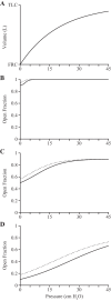
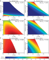
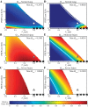
Managing patients with acute respiratory distress syndrome (ARDS) requires mechanical ventilation that balances the competing goals of sustaining life while avoiding ventilator-induced lung injury (VILI). In particular, it is reasonable to suppose that for any given ARDS patient, there must exist an optimum pair of values for tidal volume (VT) and positive end-expiratory pressure (PEEP) that together minimize the risk for VILI. To find these optimum values, and thus develop a personalized approach to mechanical ventilation in ARDS, we need to be able to predict how injurious a given ventilation regimen will be in any given patient so that the minimally injurious regimen for that patient can be determined. Our goal in the present study was therefore to develop a simple computational model of the mechanical behavior of the injured lung in order to calculate potential injury cost functions to serve as predictors of VILI. We set the model parameters to represent normal, mildly injured, and severely injured lungs and estimated the amount of volutrauma and atelectrauma caused by ventilating these lungs with a range of VT and PEEP. We estimated total VILI in two ways: 1) as the sum of the contributions from volutrauma and atelectrauma and 2) as the product of their contributions. We found the product provided estimates of VILI that are more in line with our previous experimental findings. This model may thus serve as the basis for the objective choice of mechanical ventilation parameters for the injured lung.
Keywords: acute respiratory distress syndrome; atelectrauma; mechanical ventilation; volutrauma.
Copyright © 2016 the American Physiological Society.
Figures


Similar articles
-
Using injury cost functions from a predictive single-compartment model to assess the severity of mechanical ventilator-induced lung injuries.J Appl Physiol (1985). 2019 Jul 1;127(1):58-70. doi: 10.1152/japplphysiol.00770.2018. Epub 2019 May 2. J Appl Physiol (1985). 2019. PMID: 31046518 Free PMC article.
-
The future of mechanical ventilation: lessons from the present and the past.Crit Care. 2017 Jul 12;21(1):183. doi: 10.1186/s13054-017-1750-x. Crit Care. 2017. PMID: 28701178 Free PMC article. Review.
-
Comparative Effects of Volutrauma and Atelectrauma on Lung Inflammation in Experimental Acute Respiratory Distress Syndrome.Crit Care Med. 2016 Sep;44(9):e854-65. doi: 10.1097/CCM.0000000000001721. Crit Care Med. 2016. PMID: 27035236 Free PMC article.
-
Ventilator-induced lung injury during controlled ventilation in patients with acute respiratory distress syndrome: less is probably better.Expert Rev Respir Med. 2018 May;12(5):403-414. doi: 10.1080/17476348.2018.1457954. Epub 2018 Mar 29. Expert Rev Respir Med. 2018. PMID: 29575957 Review.
-
Increasing the inspiratory time and I:E ratio during mechanical ventilation aggravates ventilator-induced lung injury in mice.Crit Care. 2015 Jan 28;19(1):23. doi: 10.1186/s13054-015-0759-2. Crit Care. 2015. PMID: 25888164 Free PMC article.
Cited by
-
Ventilator-induced lung injury and lung mechanics.Ann Transl Med. 2018 Oct;6(19):378. doi: 10.21037/atm.2018.06.29. Ann Transl Med. 2018. PMID: 30460252 Free PMC article. Review.
-
Static Stretch Increases the Pro-Inflammatory Response of Rat Type 2 Alveolar Epithelial Cells to Dynamic Stretch.Front Physiol. 2022 Apr 11;13:838834. doi: 10.3389/fphys.2022.838834. eCollection 2022. Front Physiol. 2022. PMID: 35480037 Free PMC article.
-
Computational lung modelling in respiratory medicine.J R Soc Interface. 2022 Jun;19(191):20220062. doi: 10.1098/rsif.2022.0062. Epub 2022 Jun 8. J R Soc Interface. 2022. PMID: 35673857 Free PMC article. Review.
-
Three Alveolar Phenotypes Govern Lung Function in Murine Ventilator-Induced Lung Injury.Front Physiol. 2020 Jun 30;11:660. doi: 10.3389/fphys.2020.00660. eCollection 2020. Front Physiol. 2020. PMID: 32695013 Free PMC article.
-
Using injury cost functions from a predictive single-compartment model to assess the severity of mechanical ventilator-induced lung injuries.J Appl Physiol (1985). 2019 Jul 1;127(1):58-70. doi: 10.1152/japplphysiol.00770.2018. Epub 2019 May 2. J Appl Physiol (1985). 2019. PMID: 31046518 Free PMC article.
References
-
- Acute Respiratory Distress Syndrome Network. Ventilation with lower tidal volumes as compared with traditional tidal volumes for acute lung injury and the acute respiratory distress syndrome. N Engl J Med 342: 1301–1308, 2000. - PubMed
-
- Albaiceta GM, Taboada F, Parra D, Luyando LH, Calvo J, Menendez R, Otero J. Tomographic study of the inflection points of the pressure-volume curve in acute lung injury. Am J Respir Crit Care Med 170: 1066–1072, 2004. - PubMed
-
- Allen G, Bates JH. Dynamic mechanical consequences of deep inflation in mice depend on type and degree of lung injury. J Appl Physiol (1985) 96: 293–300, 2004. - PubMed
-
- Allen GB, Leclair T, Cloutier M, Thompson-Figueroa J, Bates JH. The response to recruitment worsens with progression of lung injury and fibrin accumulation in a mouse model of acid aspiration. Am J Physiol Lung Cell Mol Physiol 292: L1580–L1589, 2007. - PubMed
MeSH terms
Grants and funding
LinkOut - more resources
Full Text Sources
Other Literature Sources

