Review
. 2016 May 13;118(10):1593-611.
doi: 10.1161/CIRCRESAHA.116.307505.

Mitochondrial Metabolism in Aging Heart

Affiliations
Review

Mitochondrial Metabolism in Aging Heart

Edward J Lesnefsky et al. Circ Res. .

Abstract

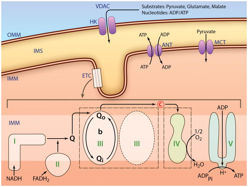
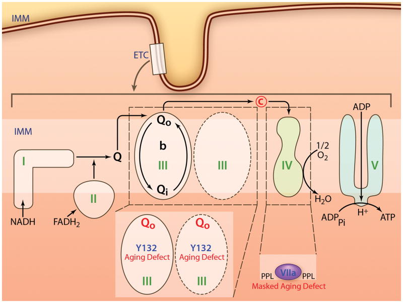
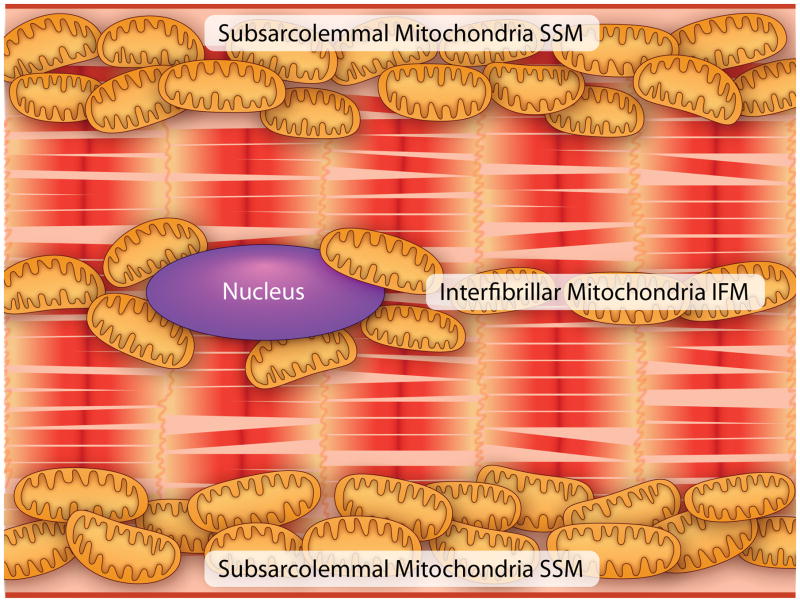
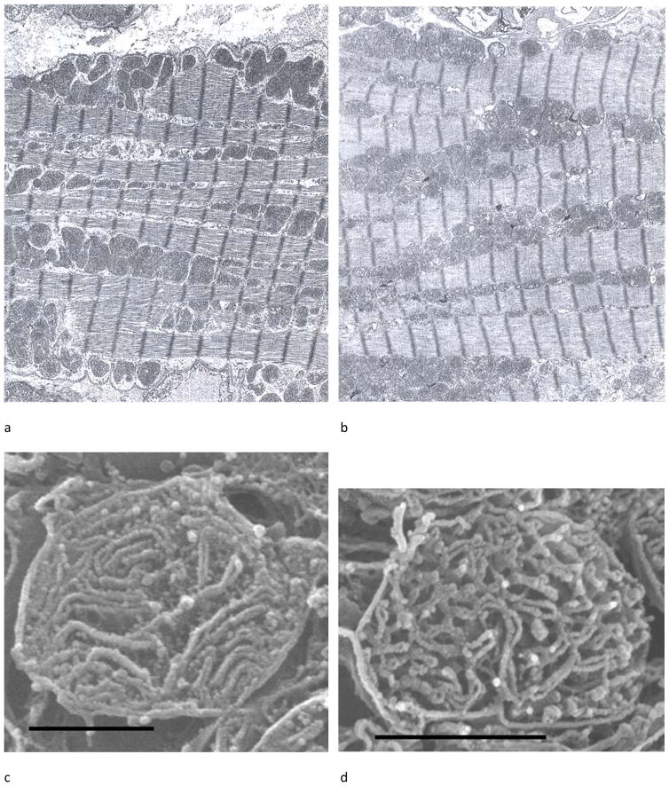
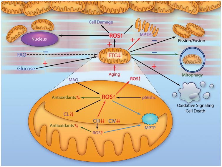
Altered mitochondrial metabolism is the underlying basis for the increased sensitivity in the aged heart to stress. The aged heart exhibits impaired metabolic flexibility, with a decreased capacity to oxidize fatty acids and enhanced dependence on glucose metabolism. Aging impairs mitochondrial oxidative phosphorylation, with a greater role played by the mitochondria located between the myofibrils, the interfibrillar mitochondria. With aging, there is a decrease in activity of complexes III and IV, which account for the decrease in respiration. Furthermore, aging decreases mitochondrial content among the myofibrils. The end result is that in the interfibrillar area, there is ≈50% decrease in mitochondrial function, affecting all substrates. The defective mitochondria persist in the aged heart, leading to enhanced oxidant production and oxidative injury and the activation of oxidant signaling for cell death. Aging defects in mitochondria represent new therapeutic targets, whether by manipulation of the mitochondrial proteome, modulation of electron transport, activation of biogenesis or mitophagy, or the regulation of mitochondrial fission and fusion. These mechanisms provide new ways to attenuate cardiac disease in elders by preemptive treatment of age-related defects, in contrast to the treatment of disease-induced dysfunction.

Keywords: cardiolipin; electron transport chain complex proteins; fatty acid oxidation complex; oxidative phosphorylation; reactive oxygen species.


Conflict of interest statement

The authors state they have no conflict of interest or financial interests to disclose.

Figures

Figure 1
Figure 1
Schematic of mitochondrial metabolism. OM, mitochondrial outer membrane; HK, hexokinase; VDAC, voltage-dependent anion channel; IMS, intermembrane space; IM, mitochondrial inner membrane; MCT, monocarboxylate transporter; ANT, adenine nucleotide translocase; IMM, mitochondrial inner membrane; ETC, electron transport chain; Q, ubiquinone; C, cytochrome c. (Illustration Credit: Ben Smith).
Figure 2
Figure 2
Schematic of the interfibrillar mitochondrial defects in the aged heart. Abbreviations similar to Figure 1. Qo site, ubiquinol binding site on cytochrome bl with proposed defect in Y132; PPL, phospholipid, proposed defect affecting subunit VIIa in complex IV. (Illustration Credit: Ben Smith).
Figure 3
Figure 3
A schematic of a cardiomyocyte to highlight the location of subsarcolemmal (SSM) and interfibrillar mitochondria (IFM). (Illustration Credit: Ben Smith).
Figure 4
Figure 4
Ultrastructure of young and aged Fischer 344 rat hearts. (a) Electron micrograph of myocardium of a 6-month rat. (b) Myocardium of a 28-month rat. SSM are clustered in groups under the sarcolemmal membrane; IFM are between the myofibrils. There are no structural differences noted between the two ages. (c) Isolated SSM from a 24-month rat, osmium-extracted. The cristae morphology is mixed between lamelliform and tubular. (d) Isolated IFM from a 24-month rat, osmium-extracted. Cristae are primarily tubular. Magnification in (a) and (b) is 6000 and the bar in (c) and (d) is 1 μm.
Figure 5
Figure 5
(a) Osmium-extracted cardiomyocyte in situ from an adult rat heart observed in cross-section by HRSEM. The sarcolemma is pointed out by the series of white arrows. The SSM are under the sarcolemma; the black arrow to Fig. 1b where a typical SSM is shown at higher magnification. The box identifies the more central area where the myofibrils have been extracted by the osmium treatment exposing the IFM. The arrow to Fig. 1c shows an IFM in higher magnification. (b) A SSM containing lamelliform cristae exclusively. (c) An IFM with tubular cristae forming a lattice. Scale line for (a) = 4 μm and for (b) and (c) = 0.5 μm. The images are taken from Figure 1a and Figure 2a and 2d from reference .
Figure 6
Figure 6
A schematic of a cardiomyocyte to show defects present in global and mitochondrial metabolism with age in concert with mechanisms of mitochondria-driven cellular injury. The aged heart exhibits impaired fatty acid oxidation (FAO) and preserved oxidation of glucose. Aging leads to defects in the electron transport chain that involve complexes III and IV. Monoamine oxidase (MAO) and p66shc are significant sources of oxidants. There is a decrease in cardiolipin (CL). Antioxidant contents are relatively unchanged, although a decrease in matrix antioxidant capacity leads to mitochondrial damage. Age-related damage enhances the production of ROS that lead to mitochondrial and cell damage. Additionally, effector mechanisms of age-induced mitochondrial damage discussed include an increased susceptibility to mitochondrial permeability transition pore opening (MPTP) (Section 6B); impaired mitochondrial dynamics favoring fusion over fission (Section 8); likely impaired mitophagy (Section 9); and enhanced oxidant derived signaling to activate cell death programs (Section 6C). (Illustration Credit: Ben Smith).

Similar articles

Cited by

References

    1. Koonen DP, Febbraio M, Bonnet S, Nagendran J, Young ME, Michelakis ED, Dyck JR. Cd36 expression contributes to age-induced cardiomyopathy in mice. Circulation. 2007;116:2139–2147. - PubMed
    1. Lesnefsky EJ, Moghaddas S, Tandler B, Kerner J, Hoppel CL. Mitochondrial dysfunction in cardiac disease: Ischemia-reperfusion, aging, and heart failure. J Mol Cell Cardiol. 2001;33:1065–1089. - PubMed
    1. Lopaschuk GD, Ussher JR, Folmes CD, Jaswal JS, Stanley WC. Myocardial fatty acid metabolism in health and disease. Physiol Rev. 2010;90:207–258. - PubMed
    1. Calvani M, Reda E, Arrigoni-Martelli E. Regulation by carnitine of myocardial fatty acid and carbohydrate metabolism under normal and pathological conditions. Basic Res Cardiol. 2000;95:75–83. - PubMed
    1. Sugden MC, Langdown ML, Harris RA, Holness MJ. Expression and regulation of pyruvate dehydrogenase kinase isoforms in the developing rat heart and in adulthood: Role of thyroid hormone status and lipid supply. Biochem J. 2000;352(Pt 3):731–738. - PMC - PubMed

Publication types

Substances