. 2016 May 13;352(6287):828-33.
doi: 10.1126/science.aae0474.

Fusion peptide of HIV-1 as a site of vulnerability to neutralizing antibody

Affiliations

Fusion peptide of HIV-1 as a site of vulnerability to neutralizing antibody

Rui Kong et al. Science. .

Abstract

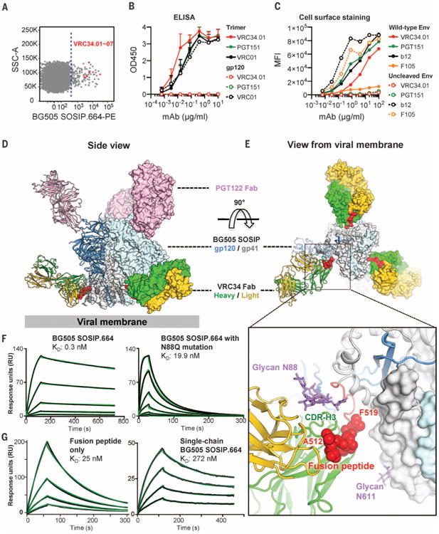
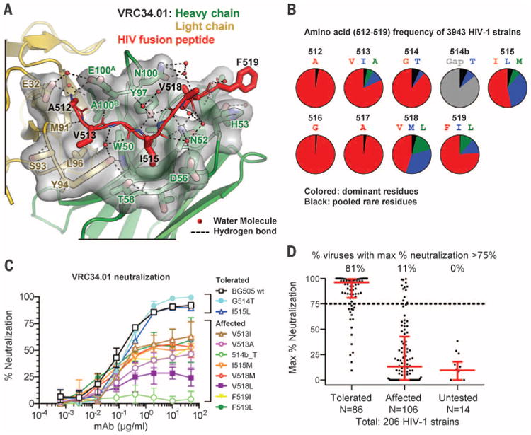
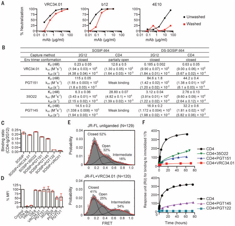
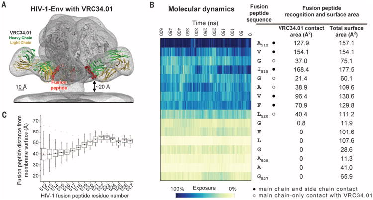
The HIV-1 fusion peptide, comprising 15 to 20 hydrophobic residues at the N terminus of the Env-gp41 subunit, is a critical component of the virus-cell entry machinery. Here, we report the identification of a neutralizing antibody, N123-VRC34.01, which targets the fusion peptide and blocks viral entry by inhibiting conformational changes in gp120 and gp41 subunits of Env required for entry. Crystal structures of N123-VRC34.01 liganded to the fusion peptide, and to the full Env trimer, revealed an epitope consisting of the N-terminal eight residues of the gp41 fusion peptide and glycan N88 of gp120, and molecular dynamics showed that the N-terminal portion of the fusion peptide can be solvent-exposed. These results reveal the fusion peptide to be a neutralizing antibody epitope and thus a target for vaccine design.


Figures

Fig. 1
Fig. 1. HIV-1-Env fusion peptide is targeted by antibody N123-VRC34.01
(A) Flow cytometric sorting of CD19+/IgG+/IgM B cells from donor N123 that bind the BG505 SOSIP.664-PE probe. Cells are shown in dot plot format, with events to the right of the dotted line sorted. Red dots indicate VRC34 lineage B cells; SSC, side scatter. (B) Antibody binding to BG505 SOSIP.664 trimer and gp120 monomer. Mean and SD of three independent experiments shown. (C) Antibody staining of 293Tcells with surface expression of wild-type or uncleaved JR-FL Env trimer. MFI, median fluorescence intensity. Data are from one of three independent experiments. (D and E) Ternary complex crystal structure comprising Env trimer (BG505 SOSIP.664) and Fabs PGT122 and VRC34.01. One Env protomer and interacting Fabs is shown in ribbon representation; the rest of the complex is shown in surface representation, with the fusion peptide highlighted in red. (D) Side view. (E) View from the viral membrane. (Inset) An expanded view of the Env-VRC34.01 interface. The fusion peptide (red) and glycan N88 (purple) comprise the VRC34.01 epitope. N-terminal residues recognized by VRC34.01 depicted as red spheres and glycan N88 as purple sticks. (F) Fab VRC34.01 binding to BG505 SOSIP.664 trimer (left panel) and to a glycan N88 knock-out mutant (N88Q, right panel) measured by SPR. Three-fold serial dilutions from 200 nM Fab injected onto the captured trimers. (G) VRC34.01 binding to fusion peptide (left panel) and to single-chain BG505 SOSIP.664 trimer (right panel), as measured by SPR. Fab VRC34.01 Fab injected in two-fold serial dilutions starting from 128 nM (left panel) and 3200 nM (right panel). For (F) and (G), three independent experiments were performed, with data from one representative experiment shown. Black lines indicate experimental data and green lines global fit to a Langmuir 1:1 binding model.
Fig. 2
Fig. 2. Fusion-peptide sequence variation is a dominant determinant of neutralization resistance to VRC34.01
(A) Crystal structure of complex between Fab VRC34.01 and a synthetic fusion peptide (comprising residues 512 through 520, red) is shown in ribbon representation with interface residues as sticks and the molecular surface of VRC34.01-peptide interactive residues in gray. Hydrogen bonds are indicated by dotted lines. The side chains of N52CDR H2 and Y97CDR H3 were hydrogen-bonded with fusion peptide residue 515 to 517, and side chains of E32CDR L1, E100ACDR H3 formed electrostatic interactions with the free amine group of residue A512, anchoring the N terminus of fusion peptide. The last residue of the synthetic fusion peptide with ordered electron density, F519, stacked with the side chain of H53CDR H2. (B) Frequency of N-terminal amino acids of the fusion peptide. Red, most prevalent; blue, second-most prevalent; green, third-most prevalent; black, pooled rare residues; gray, sequence gap. (C) VRC34.01 neutralization of BG505 Env pseudoviruses containing fusion-peptide mutations. Variants displaying more than 75% neutralization at highest antibody concentration were termed “tolerated”; variants showing less than 75% neutralization were termed “affected.” Mean and SD of three independent experiments are shown. (D) Maximum percentage neutralization by VRC34.01 against 206 HIV-1 Env pseudoviruses grouped according to their fusion-peptide sequences (25). Each dot represented one Env pseudovirus. Results were based on serial dilutions, starting at 50 μg/ml of VRC34.01. For each group, the percentage of viruses with a maximum neutralization greater than 75% is indicated on top, with the bar showing the median and interquartile range in red.
Fig. 3
Fig. 3. VRC34.01 engages prefusion closed HIV-1 Env and inhibits CD4-induced rearrangements
(A) Neutralization of JR-FL by indicated antibodies in which parallel virus-antibody mixtures were either pelleted and unbound antibody removed (washed, red lines) or not pelleted and antibody not removed (unwashed, black lines) before infection of TZM-bl cells. Representative data from one of three independent experiments are shown. The mean and SD inhibitory concentration (IC50 and IC80) values are shown in table S3. (B) Affinity of interaction of indicated antibodies with BG505 SOSIP.664 (10) or stabilized DS-SOSIP.664 (7). Standard errors from global fit of sensorgrams, obtained from independent injections of different concentrations of analyte, to a 1:1 Langmuir binding model are reported. Data sets are representative of at least two independent experiments. (C) Binding of BG505 SOSIP.664 to CD4, either alone or in the presence of antibodies (36). Mean and SD of three independent experiments are shown. (D) Influence of antibody on virus-cell attachment (37). Mean % MFI and SD of four independent experiments are shown. (E) smFRETanalysis of VRC34.01 incubation with JR-FL Env on native virions shows an increase in the high-FRET state, which is associated mechanistically with a required intermediate in the HIV-1 entry pathway (24, 38). N is the number of FRET traces analyzed. (F) Influence of antibody on the CD4-induced activation of BG505 SOSIP.664. Activation of SOSIP was monitored by assessing binding to the CD4-induced antibody, 17b, at different time points (39). Representative data of three independent experiments are shown.
Fig. 4
Fig. 4. Fusion peptide accessibility and position relative to viral membrane
(A) Superposed EM map of the BG505 SOSIP.664-VRC34.01 complex and that of HIV-1 Env trimers reconstructed from their membrane-bound context (EMDB 5019 and 5022) (gray meshes). Fab VRC34.01 bound to BG505 SOSIP.664, as extracted from the ternary complex crystal structure, was docked and shown in ribbon representation with VRC34.01-contacting fusion peptide (512 to 519) in red spheres. (B) Fusion peptide in the context of a molecular dynamic simulation of fully glycosylated HIV-1 Env trimer. The degree of solvent exposure of each residue in the fusion peptide is depicted on a blue-to-yellow color scale, with antibody-contact surface and total surface area shown for context. (C) Range of distance to the viral membrane for each residue of the HIV-1 fusion peptide over the course of the molecular dynamics simulation. The median distance for each residue is represented as a horizontal line in each box. The mean value is represented as a small filled box. The height of each box is set by the 25th and 75th interquartile range, and the whiskers are determined by the 5th and 95th percentiles. The minimum and maximum distances for each residue over the length of the simulation is represented as “x.” See fig. S23 for an analysis of immune recognition of fusion peptide in other type 1 viral fusion machines.

Similar articles

Cited by

References

    1. Colman PM, Lawrence MC. Nat Rev Mol Cell Biol. 2003;4:309–319. - PubMed
    1. Eckert DM, Kim PS. Annu Rev Biochem. 2001;70:777–810. - PubMed
    1. Harrison SC. Nat Struct Mol Biol. 2008;15:690–698. - PMC - PubMed
    1. Julien JP, et al. Science. 2013;342:1477–1483. - PMC - PubMed
    1. Lyumkis D, et al. Science. 2013;342:1484–1490. - PMC - PubMed

Publication types

MeSH terms