

Short Term Reproducibility of a High Contrast 3-D Isotropic Optic Nerve Imaging Sequence in Healthy Controls
- PMID: 27175048
- PMCID: PMC4862660
- DOI: 10.1117/12.2216834
Short Term Reproducibility of a High Contrast 3-D Isotropic Optic Nerve Imaging Sequence in Healthy Controls
Abstract
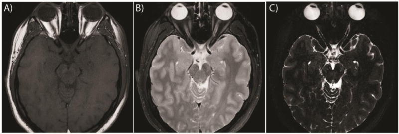
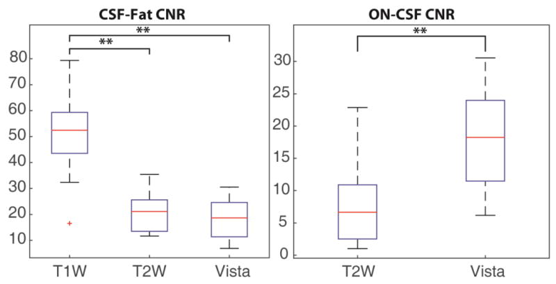
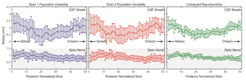
The optic nerve (ON) plays a crucial role in human vision transporting all visual information from the retina to the brain for higher order processing. There are many diseases that affect the ON structure such as optic neuritis, anterior ischemic optic neuropathy and multiple sclerosis. Because the ON is the sole pathway for visual information from the retina to areas of higher level processing, measures of ON damage have been shown to correlate well with visual deficits. Increased intracranial pressure has been shown to correlate with the size of the cerebrospinal fluid (CSF) surrounding the ON. These measures are generally taken at an arbitrary point along the nerve and do not account for changes along the length of the ON. We propose a high contrast and high-resolution 3-D acquired isotropic imaging sequence optimized for ON imaging. We have acquired scan-rescan data using the optimized sequence and a current standard of care protocol for 10 subjects. We show that this sequence has superior contrast-to-noise ratio to the current standard of care while achieving a factor of 11 higher resolution. We apply a previously published automatic pipeline to segment the ON and CSF sheath and measure the size of each individually. We show that these measures of ON size have lower short-term reproducibility than the population variance and the variability along the length of the nerve. We find that the proposed imaging protocol is (1) useful in detecting population differences and local changes and (2) a promising tool for investigating biomarkers related to structural changes of the ON.
Keywords: Magnetic Resonance Imaging; Optic Nerve; Reproducibility.
Figures
Similar articles
-
Improved Automatic Optic Nerve Radius Estimation from High Resolution MRI.Proc SPIE Int Soc Opt Eng. 2017 Feb 11;10133:101331C. doi: 10.1117/12.2254370. Epub 2017 Feb 24. Proc SPIE Int Soc Opt Eng. 2017. PMID: 28736469 Free PMC article.
-
Quantifying visual pathway axonal and myelin loss in multiple sclerosis and neuromyelitis optica.Neuroimage Clin. 2016 May 26;11:743-750. doi: 10.1016/j.nicl.2016.05.014. eCollection 2016. Neuroimage Clin. 2016. PMID: 27330974 Free PMC article.
-
Constructing a statistical atlas of the radii of the optic nerve and cerebrospinal fluid sheath in young healthy adults.Proc SPIE Int Soc Opt Eng. 2015 Mar 20;9413:941303. doi: 10.1117/12.2081887. Proc SPIE Int Soc Opt Eng. 2015. PMID: 25914505 Free PMC article.
-
Optic nerve imaging in multiple sclerosis.J Neuroimaging. 2007 Apr;17 Suppl 1:42S-45S. doi: 10.1111/j.1552-6569.2007.00136.x. J Neuroimaging. 2007. PMID: 17425734 Review.
-
Optical Coherence Tomography and Magnetic Resonance Imaging in Multiple Sclerosis and Neuromyelitis Optica Spectrum Disorder.Int J Mol Sci. 2016 Nov 15;17(11):1894. doi: 10.3390/ijms17111894. Int J Mol Sci. 2016. PMID: 27854301 Free PMC article. Review.
Cited by
-
Simultaneous total intracranial volume and posterior fossa volume estimation using multi-atlas label fusion.Hum Brain Mapp. 2017 Feb;38(2):599-616. doi: 10.1002/hbm.23432. Epub 2016 Oct 11. Hum Brain Mapp. 2017. PMID: 27726243 Free PMC article.
-
The Evaluation of Optic Nerves Using 7 Tesla "Silent" Zero Echo Time Imaging in Patients with Leber's Hereditary Optic Neuropathy with or without Idebenone Treatment.J Clin Med. 2020 Apr 13;9(4):1112. doi: 10.3390/jcm9041112. J Clin Med. 2020. PMID: 32295018 Free PMC article.
-
Quantitative characterization of optic nerve atrophy in patients with multiple sclerosis.Mult Scler J Exp Transl Clin. 2017 Sep 13;3(3):2055217317730097. doi: 10.1177/2055217317730097. eCollection 2017 Jul-Sep. Mult Scler J Exp Transl Clin. 2017. PMID: 28932410 Free PMC article.
-
Evaluating the Utility of a Postprocessing Algorithm for MRI Evaluation of Optic Neuritis.AJNR Am J Neuroradiol. 2019 Jun;40(6):1043-1048. doi: 10.3174/ajnr.A6057. Epub 2019 May 2. AJNR Am J Neuroradiol. 2019. PMID: 31048299 Free PMC article.
References
-
- O.N.S. Group. Visual function 15 years after optic neuritis: a final follow-up report from the Optic Neuritis Treatment Trial. Ophthalmology. 2008;115(6):1079. - PubMed
-
- Rizzo JF, III, Andreoli CM, Rabinov JD. Use of magnetic resonance imaging to differentiate optic neuritis and nonarteritic anterior ischemic optic neuropathy. Ophthalmology. 2002;109(9):1679–1684. - PubMed
-
- Toosy AT, Mason DF, Miller DH. Optic neuritis. The Lancet Neurology. 2014;13(1):83–99. - PubMed
Grants and funding
LinkOut - more resources
Full Text Sources
Other Literature Sources
Miscellaneous