

Paracrine regulation of matrix metalloproteinases contributes to cancer cell invasion by hepatocellular carcinoma-secreted 14-3-3σ
- PMID: 27175590
- PMCID: PMC5095053
- DOI: 10.18632/oncotarget.9234
Paracrine regulation of matrix metalloproteinases contributes to cancer cell invasion by hepatocellular carcinoma-secreted 14-3-3σ
Abstract
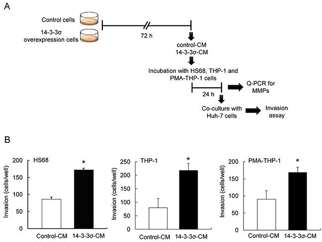
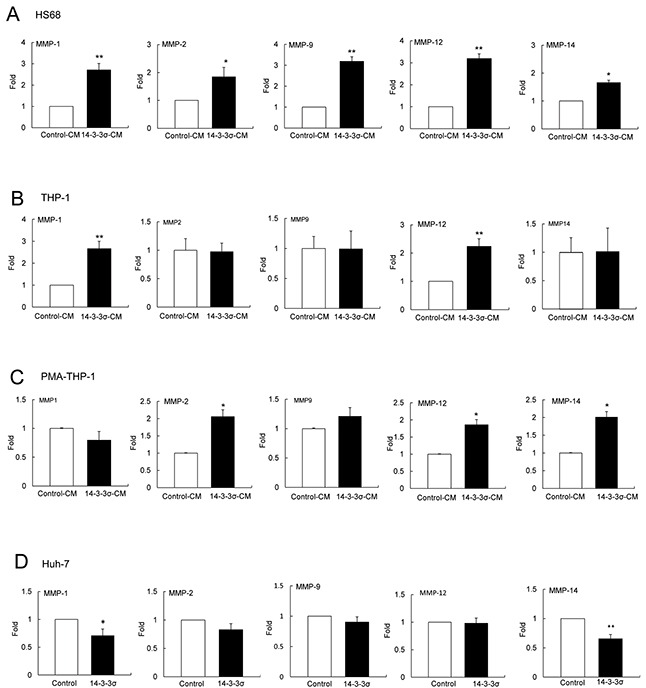
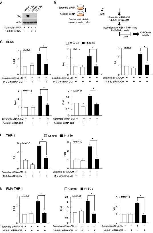
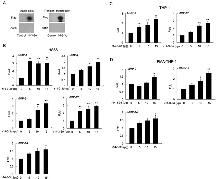
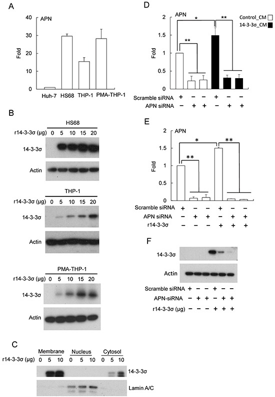
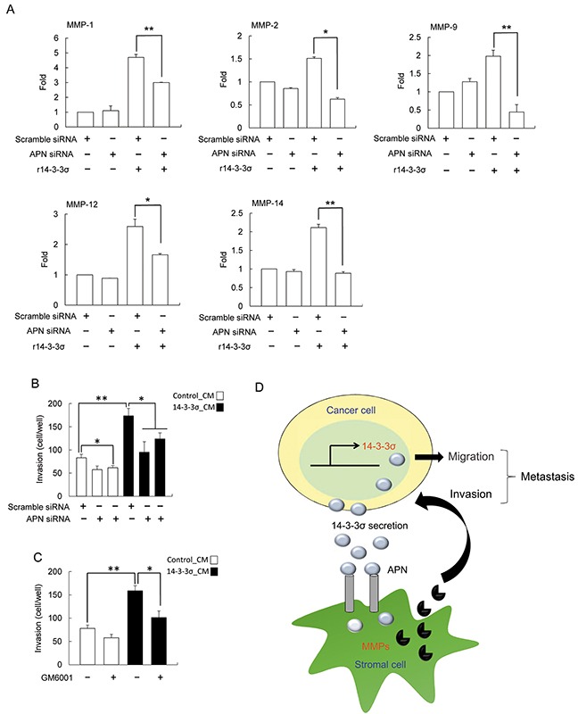
14-3-3σ overexpression results in enhanced hepatocellular carcinoma (HCC) cell migration and HCC tumor vascular-invasion is significantly associated with 14-3-3σ expression. However, increased expression of 14-3-3σ paradoxically suppresses in vitro cell invasion of HCC. We hypothesize that surrounding tumor-associated stromal cells play a crucial role in 14-3-3σ-regulated HCC cell invasion. In this study, H68 fibroblasts, THP-1 and phorbol-12-myristate-13-acetate (PMA)-treated THP-1 (PMA-THP-1) cells were incubated with conditioned media of control (control-CM) and 14-3-3σ-overepxressing cells (14-3-3σ-CM), followed by co-culture with HCC cells. Invasiveness of HCC cells was examined by a Boyden chamber assay. HCC cells co-cultured with 14-3-3σ-CM treated cells significantly enhanced their invasive ability compared with control-CM treated cells. Moreover, incubation with 14-3-3σ-CM induced differential expression profiles of matrix metalloproteinases (MMPs) in fibroblasts (MMP-1, MMP-2, MMP-9, MMP-12 and MMP-14), THP-1 (MMP-1 and MMP-12) and PMA-THP-1 cells (MMP-2, MMP-12 and MMP-14). In contrast, silencing of 14-3-3σ by siRNA significantly abolished 14-3-3σ-CM induced MMPs. In addition, treatment with recombinant 14-3-3σ (r14-3-3σ) protein exhibits a similar expression profile of MMPs induced by 14-3-3σ-CM in fibroblasts, THP-1 and PMA-THP-1 cells. Finally, knockdown of aminopeptidase N (APN) significantly abrogated r14-3-3σ induced expression of MMPs in HS68 fibroblasts. These results suggest that HCC-secreted 14-3-3σ promotes expression of MMPs in cancerous surrounding cells via an APN dependent mechanism. 14-3-3σ has a paracrine effect in educating stromal cells in tumor-associated microenvironment.
Keywords: 14-3-3σ; hepatocellular carcinoma; invasion; matrix metalloproteinase; paracrine.
Conflict of interest statement
The authors declare no conflict of interest.
Figures
Similar articles
-
Fibulin-5 inhibits hepatocellular carcinoma cell migration and invasion by down-regulating matrix metalloproteinase-7 expression.BMC Cancer. 2014 Dec 12;14:938. doi: 10.1186/1471-2407-14-938. BMC Cancer. 2014. PMID: 25494879 Free PMC article.
-
14-3-3σ induces heat shock protein 70 expression in hepatocellular carcinoma.BMC Cancer. 2014 Jun 12;14:425. doi: 10.1186/1471-2407-14-425. BMC Cancer. 2014. PMID: 24923353 Free PMC article.
-
S100A4 regulates migration and invasion in hepatocellular carcinoma HepG2 cells via NF-κB-dependent MMP-9 signal.Eur Rev Med Pharmacol Sci. 2013 Sep;17(17):2372-82. Eur Rev Med Pharmacol Sci. 2013. PMID: 24065232 Clinical Trial.
-
The Role of Matrix Metalloproteinases in the Epithelial-Mesenchymal Transition of Hepatocellular Carcinoma.Anal Cell Pathol (Amst). 2019 Nov 26;2019:9423907. doi: 10.1155/2019/9423907. eCollection 2019. Anal Cell Pathol (Amst). 2019. PMID: 31886121 Free PMC article. Review.
-
Novel strategies for hepatocellular carcinoma based on MMPs science.Anticancer Agents Med Chem. 2012 Sep;12(7):753-63. doi: 10.2174/187152012802650165. Anticancer Agents Med Chem. 2012. PMID: 22292750 Review.
Cited by
-
Type IV Collagen in Human Colorectal Liver Metastases-Cellular Origin and a Circulating Biomarker.Cancers (Basel). 2022 Jul 13;14(14):3396. doi: 10.3390/cancers14143396. Cancers (Basel). 2022. PMID: 35884455 Free PMC article.
-
Integrative Analysis of Epigenome and Transcriptome Data Reveals Aberrantly Methylated Promoters and Enhancers in Hepatocellular Carcinoma.Front Oncol. 2021 Nov 10;11:769390. doi: 10.3389/fonc.2021.769390. eCollection 2021. Front Oncol. 2021. PMID: 34858848 Free PMC article.
-
Optimizing the Diagnostic Role of Alpha-Fetoprotein and Abdominal Ultrasound by Adding Overexpressed Blood mRNA Matrix Metalloproteinase-12 for Diagnosis of HCV-Related Hepatocellular Carcinoma.Gastrointest Tumors. 2019 Feb;5(3-4):100-108. doi: 10.1159/000495838. Epub 2019 Jan 9. Gastrointest Tumors. 2019. PMID: 30976581 Free PMC article.
-
Effect of survivin downregulation by simvastatin on the growth and invasion of salivary adenoid cystic carcinoma.Mol Med Rep. 2018 Aug;18(2):1939-1946. doi: 10.3892/mmr.2018.9204. Epub 2018 Jun 22. Mol Med Rep. 2018. PMID: 29956779 Free PMC article.
-
Screening of tumor grade-related mRNAs and lncRNAs for Esophagus Squamous Cell Carcinoma.J Clin Lab Anal. 2021 Jun;35(6):e23797. doi: 10.1002/jcla.23797. Epub 2021 May 7. J Clin Lab Anal. 2021. PMID: 33960436 Free PMC article.
References
-
- Solinas G, Germano G, Mantovani A, Allavena P. Tumor-associated macrophages (TAM) as major players of the cancer-related inflammation. J Leukoc. Biol. 2009;86:1065–1073. - PubMed
-
- Sica A. Role of tumor-associated macrophages in cnacer-related inflammation. Exp Oncol. 2010;32:153–158. - PubMed
-
- Casazza A, Di Conza G, Wenes M, Finisguerra V, Deschoemaeker S, Mazzone M. Tumor stroma: a complexity dicated by the hypoxic tumor microenvironment. Oncogene. 2014;33:1743–1754. - PubMed
-
- Wu SD, Ma YS, Fang Y, Liu LL, Fu D, Shen XZ. Role of the microenvironment in hepatocellular carcinoma development and progression. Cancer Treat Rev. 2012;38:218–225. - PubMed
MeSH terms
Substances
LinkOut - more resources
Full Text Sources
Other Literature Sources
Medical
Research Materials
Miscellaneous
