

Down-regulation of microRNA-320 suppresses cardiomyocyte apoptosis and protects against myocardial ischemia and reperfusion injury by targeting IGF-1
- PMID: 27175593
- PMCID: PMC5129967
- DOI: 10.18632/oncotarget.9240
Down-regulation of microRNA-320 suppresses cardiomyocyte apoptosis and protects against myocardial ischemia and reperfusion injury by targeting IGF-1
Retraction in
-
Retraction: Down-regulation of microRNA-320 suppresses cardiomyocyte apoptosis and protects against myocardial ischemia and reperfusion injury by targeting IGF-1.Oncotarget. 2025 Mar 13;16:167. doi: 10.18632/oncotarget.28704. Oncotarget. 2025. PMID: 40079919 Free PMC article. No abstract available.
Abstract
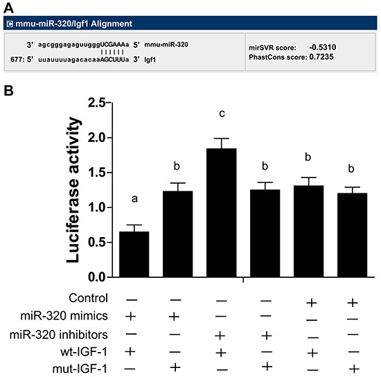
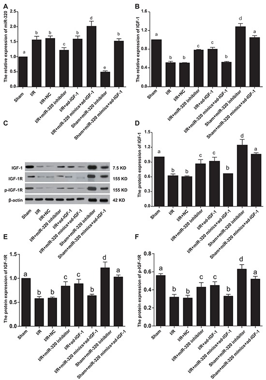
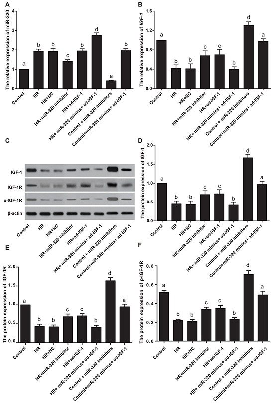
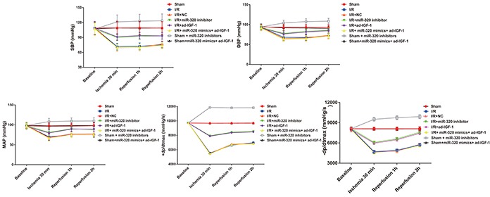
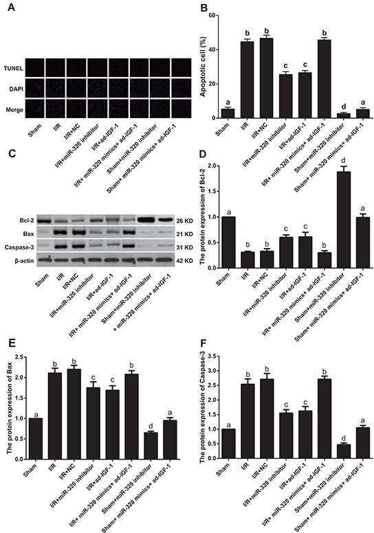
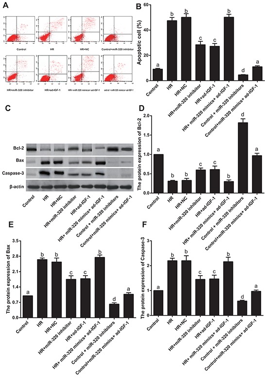
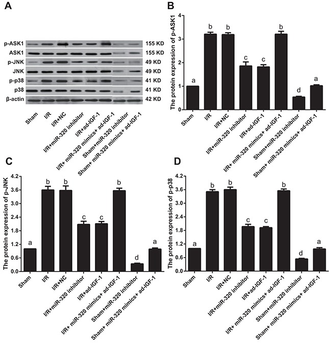
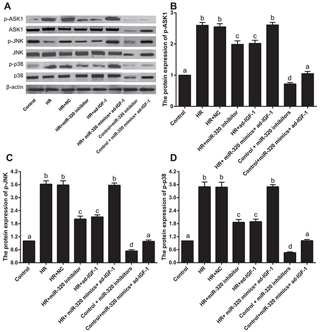
Insulin-like growth factor-1 (IGF-1) is an important regulator of cardiomyocyte homeostasis and cardiac structure, and the prosurvival and antiapoptotic effects of IGF-1 have been investigated. However, the effect of microRNA-320 (miR-320) in ischemia and reperfusion (I/R) by targeting IGF-1 is rarely discussed. We investigated the role of miR-320 in I/R injury. A total of 192 healthy female Wistar rats were divided into eight groups (n = 24). Rat heart I/R model was established. Hemodynamics, infarct size weight (ISW), heart function, and rat cardiomyocyte apoptosis were measured. Hypoxia-reoxygenation (H/R) in rat cardiomyocyte was used to simulate the I/R process. The mRNA levels of miR-320 and IGF-1, and proteins levels of IGF-1, IGF-1R, p-IGF-1R, p-ASK1, p-JNK, p-p38, Bcl-2, Bax and Caspase-3 were measured. In vivo inhibition of miR-320 expression significantly increased IGF-1 and IGF-1R mRNA levels, elevated the absolute values of SBP, DBP, MAP, ± dp/dtmax, LVEF and LVFS, decreased ISW, LVESD and LVEDd and the number of TUNEL positive cells, lowered the levels of p-ASK1, p-JNK, p-p38, Bax and Caspase-3 and increased expression of Bcl-2 compared to the I/R + NC group. Compared to H/R + NC group in vitro, miR-320 inhibition increased IGF-1 mRNA levels, inhibited cardiomyocyte apoptosis, down-regulated p-ASK, p-JNK, p-p38, Bax and Caspase-3 levels, and up-regulated Bcl-2 level. MiR-320 inhibition target elevated IGF-1 mRNA and protein levels, suppress early cardiomyocyte apoptosis of I/R, and inhibited ASK1-JNK/p38 pathway, which provides a new target for clinical study of I/R injury.
Keywords: apoptosis rate; cardiomyocytes; hypoxia-reoxygenation; ischemia-reperfusion; microRNA-320.
Conflict of interest statement
No authors in our study have any conflict of interest to disclose.
Figures
Similar articles
-
Inhibition of miR-148b ameliorates myocardial ischemia/reperfusion injury via regulation of Wnt/β-catenin signaling pathway.J Cell Physiol. 2019 Aug;234(10):17757-17766. doi: 10.1002/jcp.28401. Epub 2019 Feb 28. J Cell Physiol. 2019. PMID: 30820984
-
Anti-apoptotic effect of microRNA-30b in early phase of rat myocardial ischemia-reperfusion injury model.J Cell Biochem. 2015 Nov;116(11):2610-9. doi: 10.1002/jcb.25208. J Cell Biochem. 2015. Retraction in: J Cell Biochem. 2022 Feb;123(2):493. doi: 10.1002/jcb.30149. PMID: 25925903 Retracted.
-
Roles of MicroRNA-195 in cardiomyocyte apoptosis induced by myocardial ischemia-reperfusion injury.J Genet. 2016 Mar;95(1):99-108. doi: 10.1007/s12041-016-0616-3. J Genet. 2016. PMID: 27019437
-
Troxerutin attenuates myocardial cell apoptosis following myocardial ischemia-reperfusion injury through inhibition of miR-146a-5p expression.J Cell Physiol. 2019 Jun;234(6):9274-9282. doi: 10.1002/jcp.27607. Epub 2018 Nov 11. J Cell Physiol. 2019. PMID: 30417352
-
Pathobiology of myocardial and cardiomyocyte injury in ischemic heart disease: Perspective from seventy years of cell injury research.Exp Mol Pathol. 2024 Dec;140:104944. doi: 10.1016/j.yexmp.2024.104944. Epub 2024 Nov 21. Exp Mol Pathol. 2024. PMID: 39577392 Review.
Cited by
-
MicroRNAs in Acute ST Elevation Myocardial Infarction-A New Tool for Diagnosis and Prognosis: Therapeutic Implications.Int J Mol Sci. 2021 Apr 30;22(9):4799. doi: 10.3390/ijms22094799. Int J Mol Sci. 2021. PMID: 33946541 Free PMC article. Review.
-
Comparative miRNA expression profile analysis of porcine ovarian follicles: new insights into the initiation mechanism of follicular atresia.Front Genet. 2023 Dec 20;14:1338411. doi: 10.3389/fgene.2023.1338411. eCollection 2023. Front Genet. 2023. PMID: 38174044 Free PMC article.
-
The Role of MicroRNAs in Myocardial Infarction: From Molecular Mechanism to Clinical Application.Int J Mol Sci. 2017 Mar 31;18(4):745. doi: 10.3390/ijms18040745. Int J Mol Sci. 2017. PMID: 28362341 Free PMC article. Review.
-
Overexpressed microRNA-506 and microRNA-124 alleviate H2O2-induced human cardiomyocyte dysfunction by targeting krüppel-like factor 4/5.Mol Med Rep. 2017 Oct;16(4):5363-5369. doi: 10.3892/mmr.2017.7243. Epub 2017 Aug 14. Mol Med Rep. 2017. PMID: 28849090 Free PMC article.
-
Cell type-specific microRNA therapies for myocardial infarction.Sci Transl Med. 2021 Feb 10;13(580):eabd0914. doi: 10.1126/scitranslmed.abd0914. Sci Transl Med. 2021. PMID: 33568517 Free PMC article. Review.
References
-
- Hwa JS, Jin YC, Lee YS, Ko YS, Kim YM, Shi LY, Kim HJ, Lee JH, Ngoc TM, Bae KH, Kim YS, Chang KC. 2-methoxycinnamaldehyde from cinnamomum cassia reduces rat myocardial ischemia and reperfusion injury in vivo due to ho-1 induction. J Ethnopharmacol. 2012;139:605–615. - PubMed
-
- Arslan F, Smeets MB, O'Neill LA, Keogh B, McGuirk P, Timmers L, Tersteeg C, Hoefer IE, Doevendans PA, Pasterkamp G, de Kleijn DP. Myocardial ischemia/reperfusion injury is mediated by leukocytic toll-like receptor-2 and reduced by systemic administration of a novel anti-toll-like receptor-2 antibody. Circulation. 2010;121:80–90. - PubMed
-
- Lai RC, Arslan F, Lee MM, Sze NS, Choo A, Chen TS, Salto-Tellez M, Timmers L, Lee CN, El Oakley RM, Pasterkamp G, de Kleijn DP, Lim SK. Exosome secreted by msc reduces myocardial ischemia/reperfusion injury. Stem Cell Res. 2010;4:214–222. - PubMed
Publication types
MeSH terms
Substances
LinkOut - more resources
Full Text Sources
Other Literature Sources
Research Materials
Miscellaneous
