. 2016 May 12;23(1):44.
doi: 10.1186/s12929-016-0262-3.

Peroxisome proliferator-activated receptor-gamma dependent pathway reduces the phosphorylation of dynamin-related protein 1 and ameliorates hippocampal injury induced by global ischemia in rats

Affiliations

Peroxisome proliferator-activated receptor-gamma dependent pathway reduces the phosphorylation of dynamin-related protein 1 and ameliorates hippocampal injury induced by global ischemia in rats

Yao-Chung Chuang et al. J Biomed Sci. .

Abstract

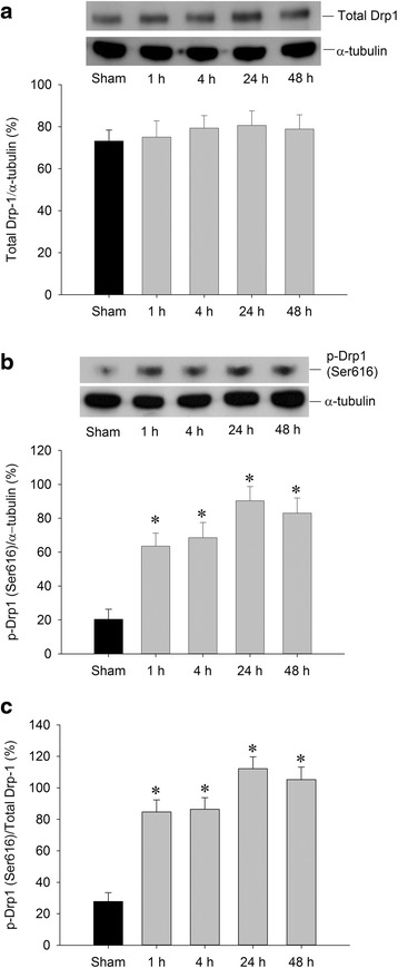
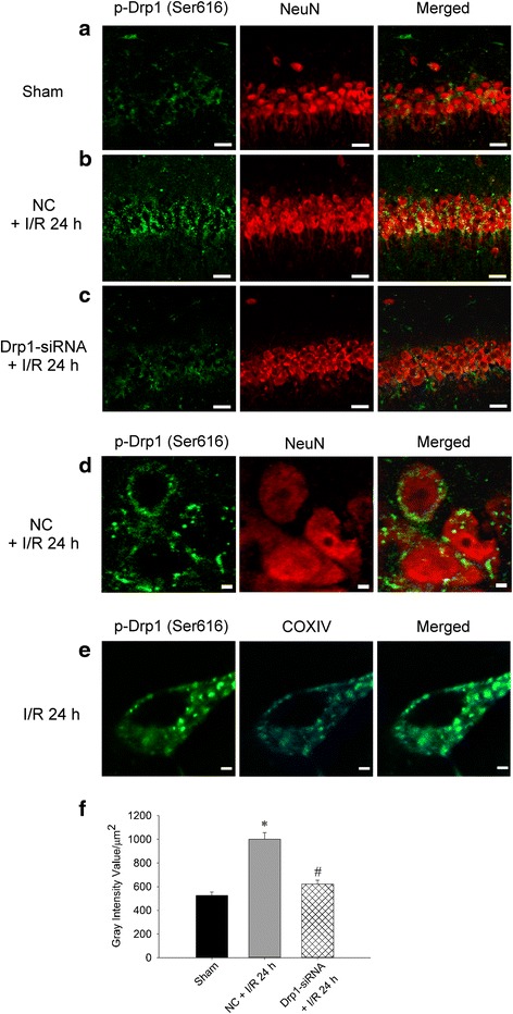
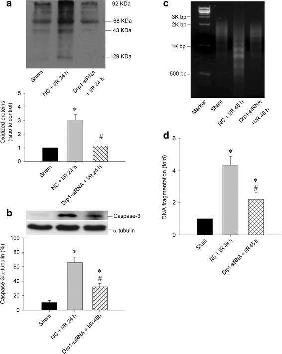
Background: Dynamin-related protein 1 (Drp1) is a mitochondrial fission protein that, upon phosphorylation at serine 616 (p-Drp1(Ser616)), plays a pivotal role in neuronal death after ischemia. In the present study, we hypothesized that peroxisome proliferator-activated receptor-gamma (PPARγ)-dependent pathway can reduce the expression of p-Drp1(Ser616) and ameliorate hippocampal injury induced by global ischemia in rats.

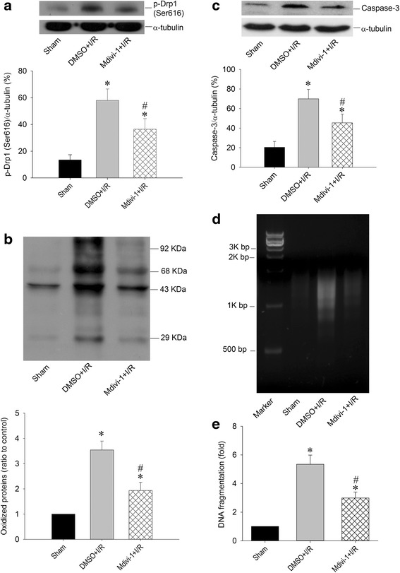
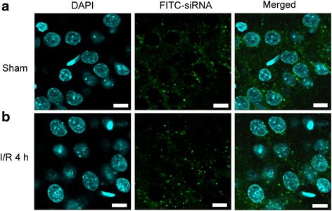
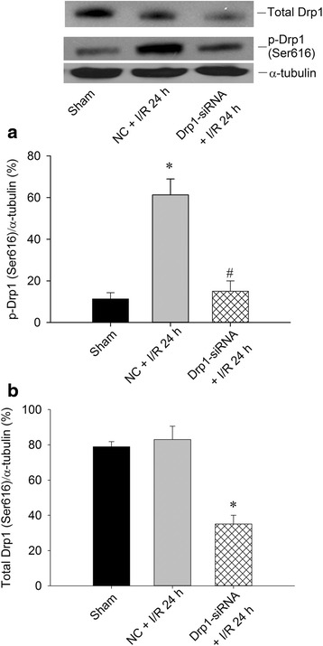
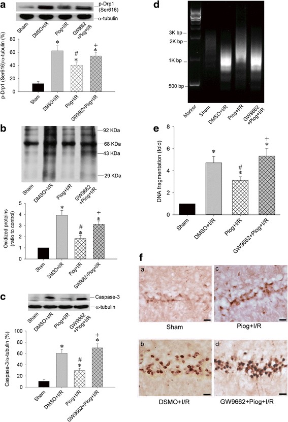
Results: We found that pretreatment of the rats with Mdivi-1, a selective Drp1 inhibitor, decreased the level of transient global ischemia (TGI)-induced p-Drp1(Ser616) and reduced cellular contents of oxidized proteins, activated caspase-3 expression as well as the extent of DNA fragmentation. Delivery of siRNA against Drp1 attenuated the expression of p-Drp1(Ser616) that was accompanied by alleviation of the TGI-induced protein oxidation, activated caspase-3 expression and DNA fragmentation in hippocampal proteins. Exogenous application of pioglitazone, a PPARγ agonist, reduced the p-Drp1(Ser616) expression, decreased TGI-induced oxidative stress and activated caspase-3 expression, lessened the extents of DNA fragmentation, and diminished the numbers of TUNEL-positive neuronal cells; all of these effects were reversed by GW9662, a PPARγ antagonist.

Conclusions: Our findings thus indicated that inhibition of TGI-induced p-Drp1(Ser616) expression by Drp1 inhibitor and Drp1-siRNA can decrease protein oxidation, activated caspase-3 expression and neuronal damage in the hippocampal CA1 subfield. PPARγ agonist, through PPARγ-dependent mechanism and via decreasing p-Drp1(Ser616) expression, can exert anti-oxidative and anti-apoptotic effects against ischemic neuronal injury.

Keywords: Apoptosis; Dynamin-related protein 1; Global ischemia; Hippocampus; Peroxisome proliferator-activated receptor-gamma; Pioglitazone.


Figures

Fig. 1
Fig. 1
Transient induction of p-Drp1(Ser616) by TGI. Hippocampal CA1 samples were collected from the rats at indicated times after 10-min TGI or sham-operated controls followed by protein extraction and western analysis for detection of total Drp1 in (a) and p-Drp1(Ser616) in (b). The same blots were also probed with α-tubulin antibody to serve as an internal reference control for equal loading of proteins in each lane. The ratio change of p-Drp1(Ser616)/total Drp was shown in (c). Values are mean ± SEM from representative blots and quantitative analyses from 4-6 animals in each experimental group are shown. *P < 0.05 versus sham control group in the Scheff′e multiple-range test
Fig. 2
Fig. 2
Mdivi-1 reduced Drp1 phosphorylation, protein oxidation, and DNA fragmentation after TGI. Rats were treated intraperitoneally with Drp1 inhibitor Mdivi-1 (2.4 mg/kg) or its solvent DMSO 30 min before TGI. Total proteins were extracted from the hippocampal CA1 subfield of sham-operated controls or treated animals 24 h after 10-min TGI for detection of p-Drp1(Ser616) in (a), protein oxidation in (b) and activated caspase-3 expression in (c). DNA was isolated from the hippocampal CA1 subfield of sham-operated controls, DMSO + I/R and Mdivi-1 + I/R 48 h after TGI for detection of DNA fragmentation by PCR assay (d), protein lysates from hippocampal CA1 tissues were collected 48 h after TGI for detection of DNA fragmentation by sandwich ELISA in (e). Values are mean ± SEM from representative blots and quantitative analysis from 5–6 animals in each experimental group (a, b and c). Values in (e) are fold changes with reference to sham-operated controls and are mean ± SEM of 4 animals in each experimental group. *P < 0.05 versus sham control group, #P < 0.05 versus DMSO + I/R group in the Scheffé multiple-range test. I/R: ischemia/reperfusion
Fig. 3
Fig. 3
Successful delivery of siRNA into hippocampal CA1 subfield. Fluorescent double staining of FITC-siRNA (green) and DAPI (blue) were observed in the hippocampal CA1 subfield 24 h after injection of 400 nl FITC-siRNA (10 μM), which was distributed in the cytosol of hippocampal CA1 in both sham-control (a) and in the rats subjected to TGI-reperfusion for 4 h (b). Scale bar: 10 μm. I/R: ischemia/reperfusion
Fig. 4
Fig. 4
Western blotting of p-Drp1(Ser616) and total Drp1 expression after Drp1-siRNA in the hippocampal CA1 subfield after TGI. After microinjection with Drp1-siRNA (0.05 nM in a total volume of 400 nl) into the CA1 subfield 24 h before TGI, total proteins were isolated from hippocampal CA1 subfield of sham-operated controls, control siRNA with TGI, or Drp1-siRNA animals after 10 min of TGI with 24 h reperfusion for detection of p-Drp1(Ser616) in (a) and total Drp1 in (b). The same blots were also probed with a α-tubulin antibody to serve as an internal control for equal loading of proteins in each lane. Values are mean ± SEM from representative blots and quantitative analysis from 4-6 animals in each experimental group. I/R: ischemia/reperfusion, NC: negative control siRNA
Fig. 5
Fig. 5
Drp1-siRNA downregulates p-Drp1(Ser616) expression in the hippocampal CA1 subfield after TGI. Fluorescent double staining of p-Drp1 (green) and NeuN (red) in the hippocampal CA1 subfield in a sham control group, b ischemia/reperfusion 24 h with negative control siRNA and c siRNA for Drp1 and ischemia/reperfusion 24 h. NeuN showed the nuclear distribution while p-Drp1 were dispersed in the cytoplasm. Scale bars, 50 μm Merged images with higher magnification demonstrate that p-Drp1(Ser616) and NeuN-positive cells localized separately in the nucleus and non-nuclear cytoplasm in neurons in (d). Scale bars, 2 μm. A semi-quantitative data about the change of p-Drp1(Ser616) expression after Drp1-siRNA for Fig. 5 a-c was shown in (f). Fluorescent double staining of p-Drp1(Ser616) (green) and COXIV (blue) in the neuron of the hippocampal CA1 subfield; merged image shows the co-localization in mitochondria in neurons under the condition of ischemia/reperfusion for 24 h (e). Scale bars, 2 μm. I/R: ischemia/reperfusion, NC: negative control siRNA. COXIV: cytochrome c oxidase subunit 4
Fig. 6
Fig. 6
Drp1 siRNA attenuates oxidative stress and decreases DNA fragmentation in hippocampal CA1 subfield after TGI. After microinjection with Drp1 siRNA (0.05 nM in a total volume of 400 nl) into the CA1 subfield 24 h before TGI, Total proteins were isolated from the hippocampal CA1 subfield of sham-operated controls, control siRNA with TGI, or Drp1 siRNA with TGI for protein oxidation in (a) and activated caspase-3 expression in (b). DNA was isolated from collected hippocampal CA1 subfield of sham-operated controls, vehicle with negative control siRNA, and Drp1-siRNA 48 h after TGI for detection of DNA fragmentation by PCR assay in (c) Hippocampal CA1 tissues were collected 48 h after TGI for detection of DNA fragmentation by sandwich ELISA in (d). Values are mean ± SEM from representative blots and quantitative analysis from 5–6 animals in each experimental group (a and b). Values are fold changes in (d) with reference to sham-control; mean ± SEM of 5–7 animals in each experimental group. *P < 0.05 vs. sham-control group and #P < 0.05 vs. negative control siRNA + I/R in the Scheff′e multiple-range test. I/R: ischemia/reperfusion, NC: negative control siRNA
Fig. 7
Fig. 7
Pioglitazone regulates Drp1 phosphorylation, protein oxidation, DNA fragmentation, and neuronal apoptosis in a PPARγ-dependant pathway after TGI. The chemical compounds microinjected into bilateral CA1 subfields as following with DMSO, pioglitazone (20 nmol) 30 min before TGI, or GW9663 (500 ng) 30 min before pioglitazone and 60 min before TGI. Total proteins were isolated from the hippocampal CA1 subfield of sham-operated controls or treated animals 24 h after 10 min of TGI for detection of p-Drp1 (Ser616) in (a) and protein oxidation in (b) and activated caspase-3 expression in (c). DNA was isolated from collected hippocampal CA1 subfield of sham-operated controls, DMSO + I/R, pioglitazone + I/R and GW9662 + pioglitazone 48 h after TGI for detection of DNA fragmentation by PCR assay (d) and hippocampal CA1 tissues were collected 48 h after TGI for detection of DNA fragmentation by sandwich ELISA in (e). Hippocampal slices were subjected to TUNEL staining to determine the extents of apoptosis in (f) which showed sham control in (a), ischemia/reperfusion with vehicle control in (b), pioglitazone with ischemia/reperfusion in (c) and GW9662 + pioglitazone and ischemia/reperfusion in (d). Values are mean ± SEM from representative blots and quantitative analyses from 5–6 animals in each experimental group (a, b and c); values in (e) are fold changes with reference to sham-control; mean ± SEM of 5-6 animals in each experimental group. *P < 0.05 vs. sham-control group, #P < 0.05 vs. DMSO + I/R and + P < 0.05 versus Piog + I/R group in the Scheff′e multiple-range test. I/R: ischemia/reperfusion. Piog: pioglitazone

References

    1. Niizuma K, Endo H, Chan PH. Oxidative stress and mitochondrial dysfunction as determinants of ischemic neuronal death and survival. J Neurochem. 2009;109(Suppl 1):133–8. doi: 10.1111/j.1471-4159.2009.05897.x. - DOI - PMC - PubMed
    1. Suen DF, Norris KL, Youle RJ. Mitochondrial dynamics and apoptosis. Genes Dev. 2008;22(12):1577–90. doi: 10.1101/gad.1658508. - DOI - PMC - PubMed
    1. Herzig S, Martinou JC. Mitochondrial dynamics: to be in good shape to survive. Curr Mol Med. 2008;8(2):131–7. doi: 10.2174/156652408783769625. - DOI - PubMed
    1. Chan DC. Mitochondrial fusion and fission in mammals. Annu Rev Cell Dev Biol. 2006;22:79–99. doi: 10.1146/annurev.cellbio.22.010305.104638. - DOI - PubMed
    1. Okamoto K, Shaw JM. Mitochondrial morphology and dynamics in yeast and multicellular eukaryotes. Annu Rev Genet. 2005;39:503–36. doi: 10.1146/annurev.genet.38.072902.093019. - DOI - PubMed