. 2016 May 12:35:78.
doi: 10.1186/s13046-016-0356-5.

PI3K p110β isoform synergizes with JNK in the regulation of glioblastoma cell proliferation and migration through Akt and FAK inhibition

Affiliations

PI3K p110β isoform synergizes with JNK in the regulation of glioblastoma cell proliferation and migration through Akt and FAK inhibition

Hua-Fu Zhao et al. J Exp Clin Cancer Res. .

Abstract

Background: Glioblastoma multiforme is the most aggressive malignant primary brain tumor, characterized by rapid growth and extensive infiltration to neighboring normal brain parenchyma. Both PI3K/Akt and JNK pathways are essential to glioblastoma cell survival, migration and invasion. Due to their hyperactivation in glioblastoma cells, PI3K and JNK are promising targets for glioblastoma treatment.

Methods: To investigate the combination effects of class IA PI3K catalytic isoforms (p110α, p110β and p110δ) and JNK inhibition on tumor cell growth and motility, glioblastoma cells and xenografts in nude mice were treated with isoform-selective PI3K inhibitors in combination with JNK inhibitor.

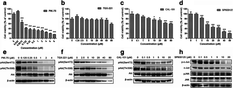
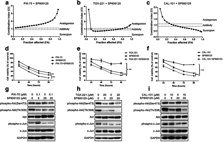
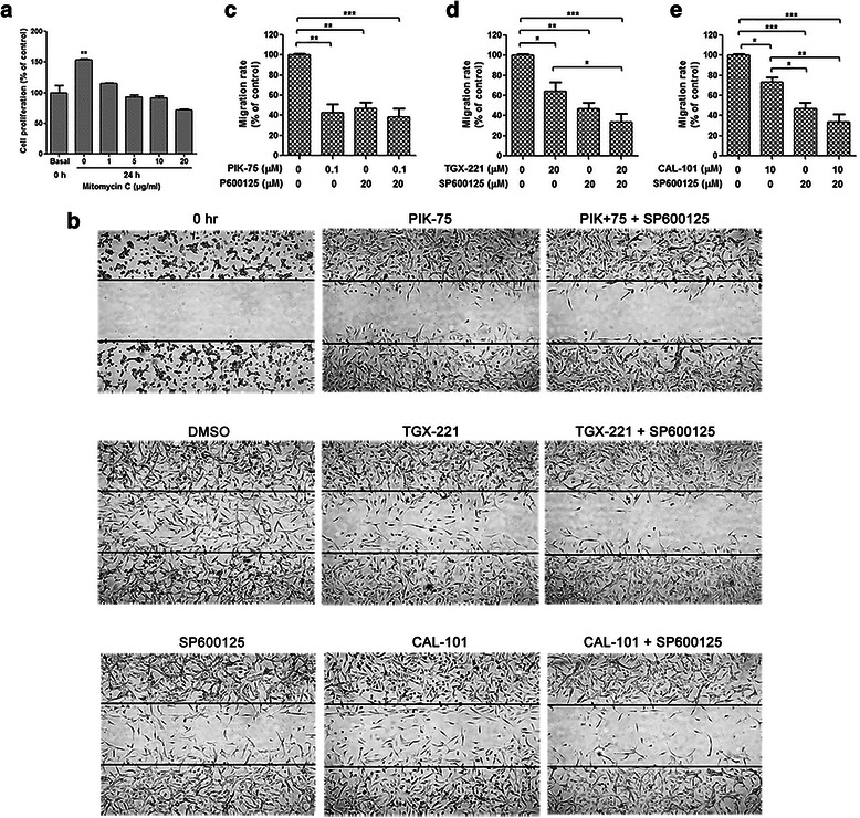
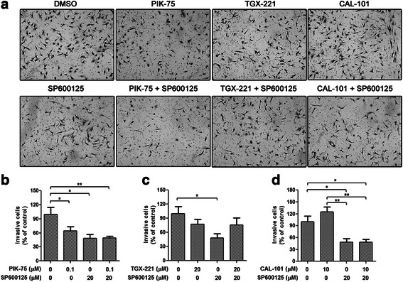
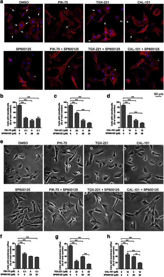
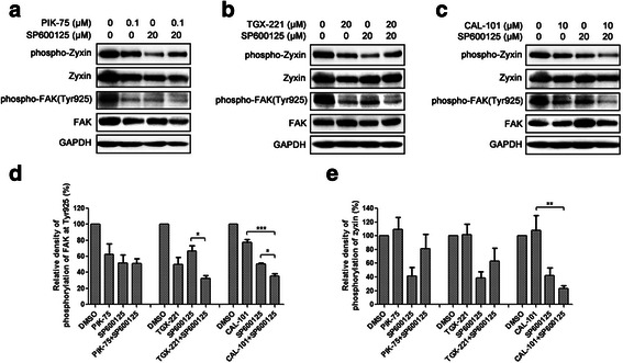
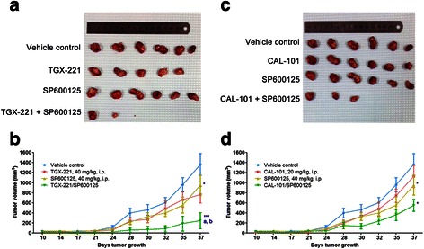
Results: We showed that combined inhibition of these PI3K isoforms and JNK exerted divergent effects on the proliferation, migration and invasion of glioblastoma cells in vitro. Pharmacological inhibition of p110β or p110δ, but not p110α, displayed synergistic inhibitory effect with JNK inhibition on glioblastoma cell proliferation and migration through decreasing phosphorylation of Akt, FAK and zyxin, leading to blockade of lamellipodia and membrane ruffles formation. No synergistic effect on invasion was observed in all the combination treatment. In vivo, combination of p110β and JNK inhibitors significantly reduced xenograft tumor growth compared with single inhibitor alone.

Conclusion: Concurrent inhibition of p110β and JNK exhibited synergistic effects on suppressing glioblastoma cell proliferation and migration in vitro and xenograft tumor growth in vivo. Our data suggest that combined inhibition of PI3K p110β isoform and JNK may serve as a potent and promising therapeutic approach for glioblastoma multiforme.

Keywords: Glioblastoma; JNK; Migration; PI3K; Proliferation; Synergism; p110β.


Figures

Fig. 1
Fig. 1
Inhibitory effects of PIK-75, TGX-221, CAL-101 and SP600125 on glioblastoma cell proliferation and signaling transduction. a-d U-87 MG cells were treated with PIK-75, TGX-221, CAL-101 or SP600125 at different concentrations for 48 h. DMSO was used as a carrier control. Cell viability of U-87 MG cells sharply decreased when treated with PIK-75, CAL-101 and SP600125, whereas TGX-221 had no significant inhibitory effect (n = 3; p values were determined by One-way ANOVA and Post Hoc multiple comparison Tukey HSD test. *: p <0.05; **: p <0.01; ***: p <0.001). e-g Isoform-selective PI3K inhibitors PIK-75, TGX-221 and CAL-101 dramatically inhibited Akt phosphorylation at Ser473 and Thr308. h JNK inhibitor SP600125 suppressed JNK signaling via reducing c-Jun phosphorylation, whereas JNK phosphorylation was not affected
Fig. 2
Fig. 2
Combination effects of isoform-selective PI3K inhibitors and SP600125 on glioblastoma cell proliferation. a-c FA-CI plots were generated using Chou-Talalay method. U-87 MG cells were treated with two inhibitors at a fixed ratio for 48 h. d-e U-87 MG cells were treated with PIK-75 (0.1 μM), TGX-221 (20 μM) or CAL-101 (10 μM) alone and combined with SP600125 (20 μM) for 24, 48, 72 and 96 h respectively. DMSO was used as a carrier control. Antagonistic effect was found in the combination of PIK-75 and SP600125, whereas SP600125 significantly synergized with TGX-221 or CAL-101 (n = 3; p values were determined by Two-way ANOVA and Post Hoc multiple comparison Tukey HSD test. ***: p <0.001). g-i U-87 MG cells were treated with PIK-75, TGX-221 or CAL-101 alone and combined with SP600125 for 3 h. Phosphorylation level of Akt was also downregulated by SP600125. Combination of TGX-211 and SP600125, as well as CAL-101 and SP600125 exhibited higher inhibition of Akt phosphorylation at Thr308
Fig. 3
Fig. 3
SP600125 potentiated inhibitory effects of isoform-selective PI3K inhibitors on glioblastoma cell migration. a Number of viable U-87 MG cells was counted using Vi-Cell Cell Viability Analyzer (Beckman Coulter) before and after treatment of mitomycin C for 24 h. Cell proliferation was inhibited by mitomycin C at 5 or 10 μg/mL without impairing viability. b Wound healing in U-87 MG cells treated with PIK-75 (0.1 μM), TGX-221 (20 μM) or CAL-101 (10 μM) alone and combined with SP600125 (20 μM) for 24 h. Cells were pretreated with 5 μg/mL of mitomycin C for 1 h. The lines indicate the edge of wound generated before drug treatment (0 h). Photographs were obtained at 50× magnification. c-e Migration rate was analyzed and expressed as the number of cells migrating into the original wounds. U-87 MG cell migration was blocked by PIK-75, TGX-221, CAL-101 and SP600125 alone. Inhibition of migration rate was reinforced by the combined treatment of TGX-221 and SP600125, as well as CAL-101 and SP600125 (n = 3; p values were determined by One-way ANOVA and Post Hoc multiple comparison Tukey HSD test. *: p <0.05; **: p <0.01; ***: p <0.001)
Fig. 4
Fig. 4
No synergistic effect on glioblastoma cell invasion was observed in the combination of isoform-selective PI3K inhibitors and SP600125. Boyden chamber invasion assay of U-87 MG cells treated with PIK-75 (0.1 μM), TGX-221 (20 μM) or CAL-101 (10 μM) alone and combined with SP600125 (20 μM) for 24 h. a Representative photographs showing the invasive cells that had passed through matrigel to the lower surface of the membrane at 100× magnification. Invaded cells from 5 representative fields were counted. b-d Number of invasive cells was significantly decreased by PIK-75 and SP600125, and all the combinations did not enhanced the inhibitory effect (n = 3; p values were determined by One-way ANOVA and Post Hoc multiple comparison Tukey HSD test. *: p <0.05; **: p <0.01)
Fig. 5
Fig. 5
SP600125 potentiated inhibitory effects of isoform-selective PI3K inhibitors on lamellipodia and membrane ruffles formation. a Immunocytochemistry of U-87 MG cells stained with Alexa Fluor 594-conjugated phalloidin (red) after treatment with PIK-75 (0.1 μM), TGX-221 (20 μM) or CAL-101 (10 μM) alone and combined with SP600125 (20 μM) for 3 h. Lamellipodia (white arrow) were decreased after inhibition of p110α, p110β, p110δ and JNK, while stress fibers (arrowhead) were widely developed at the presence of SP600125. Bar = 50 μm. b-d Cells with lamellipodia were counted and data were expressed as the ratio of number of cells with lamellipodia over total cell number. SP600125 potentiated the inhibitory effect of TGX-221 and CAL-101 on lamellipodia formation (n = 3; p values were determined by One-way ANOVA and Post Hoc multiple comparison Tukey HSD test. ***: p <0.001). e Phase contrast microscopy of U-87 MG cells after treatment with PIK-75, TGX-221 or CAL-101 alone and combined with SP600125 for 3 h. Cells with membrane ruffles (black arrow) were reduced by the treatment of isoform-selective PI3K inhibitors and SP600125. Photographs were obtained at 200× magnification. f-h Cells with membrane ruffles were counted and data were expressed as the ratio of number of cells with membrane ruffles over total cell number. SP600125 also enhanced the inhibitory effect of TGX-221 and CAL-101 on membrane ruffles formation (n = 3; p values were determined by One-way ANOVA and Post Hoc multiple comparison Tukey HSD test. **: p <0.01; ***: p <0.001)
Fig. 6
Fig. 6
Isoform-selective PI3K inhibitors and SP600125 affected focal adhesion and cytoskeleton-related signaling. a-c U-87 MG cells were treated with PIK-75 (0.1 μM), TGX-221 (20 μM) or CAL-101 (10 μM) alone and combined with SP600125 (20 μM) for 24 h. Whole cell lysates were separated by SDA-PAGE, and the phosphorylated and total protein levels were determined by Western blotting using antibodies against the corresponding phosphorylated and total proteins. Expression of GAPDH was served as a loading control. Data were representative of three independent experiments. Density of FAK d and zyxin e phosphorylation levels were relative to total FAK and zyxin protein expression levels. Combination of TGX-221 and SP600125 showed a higher inhibition of FAK phosphorylation, while combination of CAL-101 and SP600125 displayed higher inhibition of FAK and zyxin phosphorylation (n = 3; p values were determined by One-way ANOVA and Post Hoc multiple comparison Tukey HSD test. *: p <0.05; **: p <0.01)
Fig. 7
Fig. 7
Combined inhibition of p110β and JNK exerted synergism to suppress xenograft tumor growth in vivo. Human glioblastoma U-87 MG cells (5 × 106 cells) were subcutaneously injected into the Balb/C nude mice. Nine days after inoculation, mice were intraperitoneally (i.p.) injected once daily for 7 days with vehicle, TGX-221 (40 mg/kg) and CAL-101 (20 mg/kg) alone, or in combination with SP600125 (40 mg/kg). Measurement of tumor volumes started on the day of the first administration. a, c Representative subcutaneous tumor xenografts from mice sacrificed after 37-day post-administration. b, d Summary data of tumor volumes from 10-day post-administration to the end of the experiment. (n = 6; p values were determined by One-way repeated measures ANOVA and Post Hoc multiple comparison Tukey HSD test. *: p <0.05; ***: p <0.001 compared with vehicle control; a: p <0.05 compared with PI3K inhibitor; b: p <0.05 compared with SP600125)

References

    1. Louis DN, Ohgaki H, Wiestler OD, Cavenee WK, Burger PC, Jouvet A, Scheithauer BW, Kleihues P. WHO classification of tumours of the central nervous system. Acta Neuropathol. 2007;2007(114):97–109. - PMC - PubMed
    1. Wang ZL, Zhang CB, Cai JQ, Li QB, Wang Z, Jiang T. Integrated analysis of genome-wide DNA methylation, gene expression and protein expression profiles in molecular subtypes of WHO II-IV gliomas. J Exp Clin Cancer Res. 2015;34:127. doi: 10.1186/s13046-015-0249-z. - DOI - PMC - PubMed
    1. Cantley LC. The phosphoinositide 3-kinase pathway. Science. 2002;296:1655–1657. doi: 10.1126/science.296.5573.1655. - DOI - PubMed
    1. Usatyuk PV, Fu P, Mohan V, Epshtein Y, Jacobson JR, Gomez-Cambronero J, Wary KK, Bindokas V, Dudek SM, Salgia R, et al. Role of c-Met/phosphatidylinositol 3-kinase (PI3k)/Akt signaling in hepatocyte growth factor (HGF)-mediated lamellipodia formation, reactive oxygen species (ROS) generation, and motility of lung endothelial cells. J Biol Chem. 2014;289:13476–91. - PMC - PubMed
    1. Sossey-Alaoui K, Li X, Ranalli TA, Cowell JK. WAVE3-mediated cell migration and lamellipodia formation are regulated downstream of phosphatidylinositol 3-kinase. J Biol Chem. 2005;280:21748–21755. doi: 10.1074/jbc.M500503200. - DOI - PubMed

Publication types

MeSH terms

Substances