

Strain and strain rate echocardiography in children with Wilson's disease
- PMID: 27176875
- PMCID: PMC5378936
- DOI: 10.5830/CVJA-2016-028
Strain and strain rate echocardiography in children with Wilson's disease
Abstract
Objective: This study aimed to evaluate strain and strain rate echocardiography in children with Wilson's disease to detect early cardiac dysfunction.
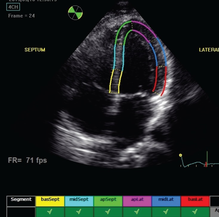
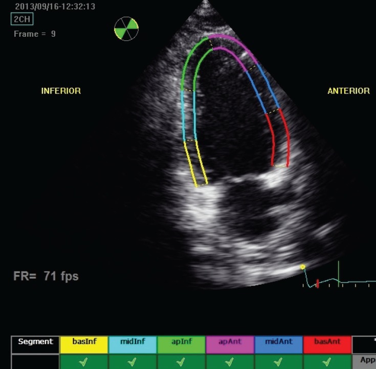
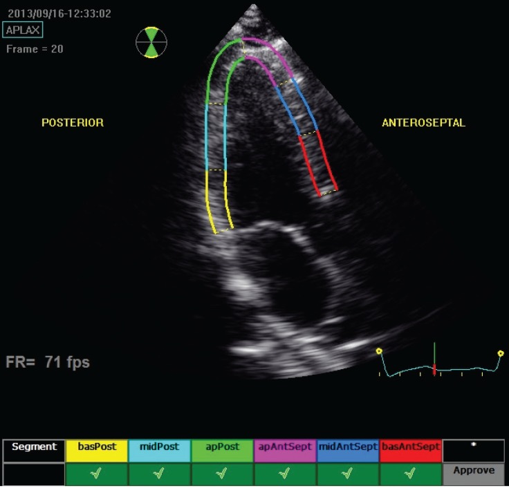
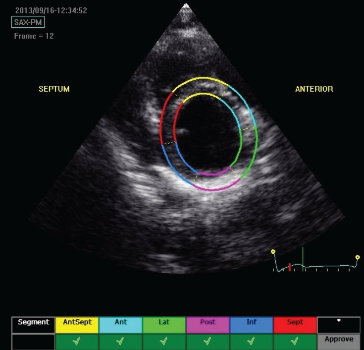
Methods: In this study, 21 patients with Wilson's disease and a control group of 20 age- and gender-matched healthy children were included. All the patients and the control group were evaluated with two-dimensional (2D) and colour-coded conventional transthoracic echocardiography by the same paediatric cardiologist using the same echocardiography machine (Vivid E9, GE Healthcare, Norway) in standard precordial positions, according to the American Society of Echocardiography recommendations. 2D strain and strain rate echocardiography were performed after the ECG probes of the echocardiography machine were adjusted for ECG monitoring. Longitudinal, transverse and radial strain, and strain rate were assessed from six basal and six mid-ventricular segments of the left ventricle, as recommended by the American Society of Echocardiography.
Results: Left ventricular wall thickness, systolic and diastolic diameters, left ventricular diameters normalised to body surface area, end-systolic and end-diastolic volumes, cardiac output and cardiac index values were within normal limits and statistically similar in the patient and control groups (p > 0.05). Global strain and strain rate: the patient group had a statistically significant lower peak A longitudinal velocity of the left basal point and peak E longitudinal velocity of the left basal (VAbasR) point, and higher global peak A longitudinal/circumferential strain rate (GSRa) compared to the corresponding values of the control group (p < 0.05). Radial strain and strain rate: end-systolic rotation [ROT (ES)] was statistically significantly lower in the patient group (p < 0.05). Longitudinal strain and strain rate: end-systolic longitudinal strain [SLSC (ES)] and positive peak transverse strain (STSR peak P) were statistically significantly lower in the patient group (p < 0.05). Segmental analysis showed that rotational strain measurement of the anterior and lateral segments of the patient group were statistically significantly lower than the corresponding values of the control group (p < 0.05). Segmental analysis showed statistically significantly lower values of endsystolic longitudinal strain [STSR (ES)] of the basal lateral (p < 0.05) and end-systolic longitudinal strain [SLSC (ES)] of the basal septal segment (p < 0.05) in the patient group. End-systolic longitudinal strain [SLSC (ES)] and positive peak transverse strain (STSR peak P) were statistically significantly lower in the patient group (p < 0.05). Segmental analysis showed statistically significantly lower values of endsystolic longitudinal strain [SLSC (ES)] of the mid-anterior and basal anterior segments (p < 0.05), end-systolic longitudinal strain [STSR (ES)] measurements of the posterior and mid-posterior segments, end-systolic longitudinal displacement [DLDC (ES)] of the basal posterior, mid-posterior and mid-antero-septal segments in the patient group.
Conclusion: Cardiac arrhythmias, cardiomyopathy and sudden cardiac death are rare complications but may be seen in children with Wilson's disease due to copper accumulation in the heart tissue. Strain and strain rate echocardiography is a relatively new and useful echocardiographic technique to evaluate cardiac function and cardiac deformation abnormalities. Our study showed that despite normal systolic function, patients with Wilson's disease showed diastolic dysfunction and regional deformation abnormalities, especially rotational strain and strain rate abnormalities.
Figures
Similar articles
-
Assessment of left ventricular systolic and diastolic abnormalities in patients with hypertrophic cardiomyopathy using real-time three-dimensional echocardiography and two-dimensional speckle tracking imaging.Cardiovasc Ultrasound. 2018 Oct 2;16(1):23. doi: 10.1186/s12947-018-0142-y. Cardiovasc Ultrasound. 2018. PMID: 30285887 Free PMC article.
-
Assessment of myocardial mechanics in patients with end-stage renal disease and renal transplant recipients using speckle tracking echocardiography.Exp Clin Transplant. 2015 Apr;13 Suppl 1:235-41. Exp Clin Transplant. 2015. PMID: 25894162
-
Ventricular septal dysfunction after surgical closure of multiple ventricular septal defects.Ann Thorac Surg. 2013 Sep;96(3):891-7. doi: 10.1016/j.athoracsur.2013.05.013. Epub 2013 Jul 26. Ann Thorac Surg. 2013. PMID: 23895887
-
Perioperative assessment of myocardial deformation.Anesth Analg. 2014 Mar;118(3):525-44. doi: 10.1213/ANE.0000000000000088. Anesth Analg. 2014. PMID: 24557101 Free PMC article. Review.
-
Left ventricular longitudinal systolic dysfunction in children with type 1 diabetes mellitus: A systematic review and meta-analysis.J Diabetes Complications. 2023 Aug;37(8):108528. doi: 10.1016/j.jdiacomp.2023.108528. Epub 2023 Jun 6. J Diabetes Complications. 2023. PMID: 37459780
Cited by
-
Left Ventricular Myocardial Deformations in Hemodialysis Children by Speckle Tracking Echocardiography.Clin Med Insights Cardiol. 2020 Jun 8;14:1179546820930015. doi: 10.1177/1179546820930015. eCollection 2020. Clin Med Insights Cardiol. 2020. PMID: 32550769 Free PMC article.
-
Cardiac assessment in Wilson's disease patients based on electrocardiography and echocardiography examination.Arch Med Sci. 2019 Jul;15(4):857-864. doi: 10.5114/aoms.2017.69728. Epub 2017 Sep 7. Arch Med Sci. 2019. PMID: 31360180 Free PMC article.
-
Assessment of biventricular systolic strain derived from the two-dimensional and three-dimensional speckle tracking echocardiography in lymphoma patients after anthracycline therapy.Int J Cardiovasc Imaging. 2017 Jun;33(6):857-868. doi: 10.1007/s10554-017-1082-6. Epub 2017 Mar 2. Int J Cardiovasc Imaging. 2017. PMID: 28255826
-
The association between systolic and diastolic dysfunction and autonomic nervous system function in children receiving chronic hemodialysis.Pediatr Nephrol. 2025 Aug;40(8):2599-2610. doi: 10.1007/s00467-024-06577-1. Epub 2025 Jan 28. Pediatr Nephrol. 2025. PMID: 39873803 Free PMC article.
References
-
- Rosencrantz R, Schilsky M. Wilson’s disease: pathogenesis and clinical considerations in diagnosis and treatment. Semin Liver Dis. 2011;31(3):245–259. - PubMed
-
- Bennett J, Hahn SH. Clinical molecular diagnosis of Wilson’s disease. Semin Liver Dis. 2011;13(3):233–238. - PubMed
-
- Aggarwal A, Bhatt M. Update on Wilson disease. Int Rev Neurobiol. 2013;110:313–348. - PubMed
-
- Kuan P. Cardiac Wilson’s disease. Chest. 1987;91(4):579–583. - PubMed
-
- Meenakshi-Sundaram S, Sinha S, Rao M, Prashanth LK, Arunodaya GR, Rao S. et al. Cardiac involvement in Wilson’s disease – an electrocardiographic observation. J Assoc Physicians India. 2004;52:294–296. - PubMed
MeSH terms
LinkOut - more resources
Full Text Sources
Other Literature Sources
Medical
