

Polarization sensitive optical coherence microscopy for brain imaging
- PMID: 27176965
- PMCID: PMC5357322
- DOI: 10.1364/OL.41.002213
Polarization sensitive optical coherence microscopy for brain imaging
Abstract
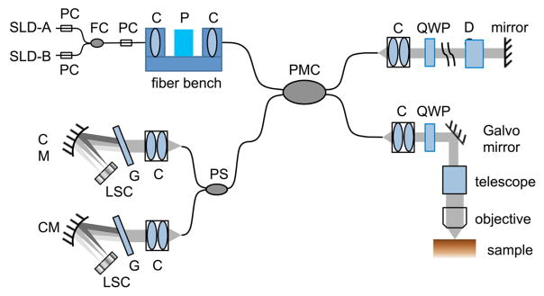
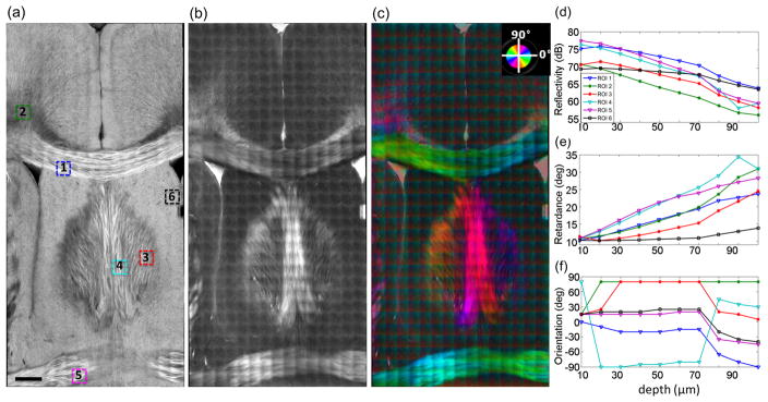
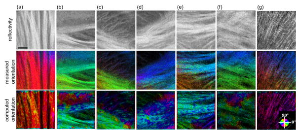
Optical coherence tomography (OCT) and optical coherence microscopy (OCM) have demonstrated the ability to investigate cyto- and myelo-architecture in the brain. Polarization-sensitive OCT provides sensitivity to additional contrast mechanisms, specifically the birefringence of myelination and, therefore, is advantageous for investigating white matter fiber tracts. In this Letter, we developed a polarization-sensitive optical coherence microscope (PS-OCM) with a 3.5 μm axial and 1.3 μm transverse resolution to investigate fiber organization and orientation at a finer scale than previously demonstrated with PS-OCT. In a reconstructed mouse brain section, we showed that at the focal depths of 20-70 μm, the PS-OCM reliably identifies the neuronal fibers and quantifies the in-plane orientation.
Figures
Similar articles
-
Determination of characteristics of degenerative joint disease using optical coherence tomography and polarization sensitive optical coherence tomography.Lasers Surg Med. 2006 Oct;38(9):852-65. doi: 10.1002/lsm.20394. Lasers Surg Med. 2006. PMID: 16998913
-
Depth-resolved birefringence and differential optical axis orientation measurements with fiber-based polarization-sensitive optical coherence tomography.Opt Lett. 2004 Sep 1;29(17):2025-7. doi: 10.1364/ol.29.002025. Opt Lett. 2004. PMID: 15455768
-
Variables affecting polarization-sensitive optical coherence tomography imaging examined through the modeling of birefringent phantoms.J Opt Soc Am A Opt Image Sci Vis. 2005 Feb;22(2):262-71. doi: 10.1364/josaa.22.000262. J Opt Soc Am A Opt Image Sci Vis. 2005. PMID: 15717555
-
Multimodal optical imaging with multiphoton microscopy and optical coherence tomography.J Biophotonics. 2012 May;5(5-6):396-403. doi: 10.1002/jbio.201100138. Epub 2012 Mar 28. J Biophotonics. 2012. PMID: 22461146 Review.
-
Optical coherence tomography for precision brain imaging, neurosurgical guidance and minimally invasive theranostics.Biosci Trends. 2018 Mar 18;12(1):12-23. doi: 10.5582/bst.2017.01258. Epub 2018 Jan 15. Biosci Trends. 2018. PMID: 29332928 Review.
Cited by
-
Advances in Diagnostic Tools and Therapeutic Approaches for Gliomas: A Comprehensive Review.Sensors (Basel). 2023 Dec 15;23(24):9842. doi: 10.3390/s23249842. Sensors (Basel). 2023. PMID: 38139688 Free PMC article. Review.
-
Robust reconstruction of local optic axis orientation with fiber-based polarization-sensitive optical coherence tomography.Biomed Opt Express. 2018 Oct 15;9(11):5437-5455. doi: 10.1364/BOE.9.005437. eCollection 2018 Nov 1. Biomed Opt Express. 2018. PMID: 30460138 Free PMC article.
-
Ultra-parallel label-free optophysiology of neural activity.iScience. 2022 Apr 27;25(5):104307. doi: 10.1016/j.isci.2022.104307. eCollection 2022 May 20. iScience. 2022. PMID: 35602935 Free PMC article.
-
Insight into the fundamental trade-offs of diffusion MRI from polarization-sensitive optical coherence tomography in ex vivo human brain.Neuroimage. 2020 Jul 1;214:116704. doi: 10.1016/j.neuroimage.2020.116704. Epub 2020 Mar 6. Neuroimage. 2020. PMID: 32151760 Free PMC article.
-
Imaging myelin degradation in ex vivo prefrontal cortex tissue blocks in Alzheimer's disease and chronic traumatic encephalopathy.Alzheimers Dement. 2025 Aug;21(8):e70582. doi: 10.1002/alz.70582. Alzheimers Dement. 2025. PMID: 40843933 Free PMC article.
References
-
- Min Eunjung, Lee Junwon, Vavilin Andrey, Jung Sunwoo, Shin Sungwon, Kim Jeehyun, Jung Woonggyu. Wide-field optical coherence microscopy of the mouse brain slice. Opt Lett. 2015;40:4420–4423. - PubMed
MeSH terms
Grants and funding
LinkOut - more resources
Full Text Sources
Other Literature Sources
