. 2016 Sep 1;23(9):1565-76.
doi: 10.1038/cdd.2016.46. Epub 2016 May 13.

RIPK3 deficiency or catalytically inactive RIPK1 provides greater benefit than MLKL deficiency in mouse models of inflammation and tissue injury

Affiliations

RIPK3 deficiency or catalytically inactive RIPK1 provides greater benefit than MLKL deficiency in mouse models of inflammation and tissue injury

K Newton et al. Cell Death Differ. .

Abstract

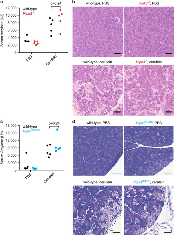
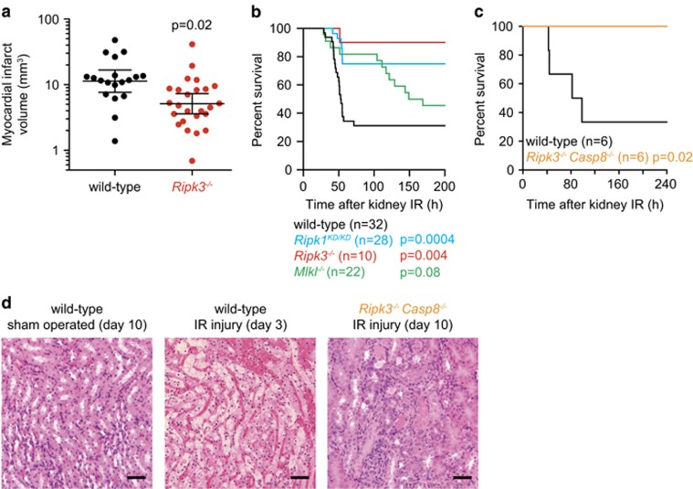
Necroptosis is a caspase-independent form of cell death that is triggered by activation of the receptor interacting serine/threonine kinase 3 (RIPK3) and phosphorylation of its pseudokinase substrate mixed lineage kinase-like (MLKL), which then translocates to membranes and promotes cell lysis. Activation of RIPK3 is regulated by the kinase RIPK1. Here we analyze the contribution of RIPK1, RIPK3, or MLKL to several mouse disease models. Loss of RIPK3 had no effect on lipopolysaccharide-induced sepsis, dextran sodium sulfate-induced colitis, cerulein-induced pancreatitis, hypoxia-induced cerebral edema, or the major cerebral artery occlusion stroke model. However, kidney ischemia-reperfusion injury, myocardial infarction, and systemic inflammation associated with A20 deficiency or high-dose tumor necrosis factor (TNF) were ameliorated by RIPK3 deficiency. Catalytically inactive RIPK1 was also beneficial in the kidney ischemia-reperfusion injury model, the high-dose TNF model, and in A20(-/-) mice. Interestingly, MLKL deficiency offered less protection in the kidney ischemia-reperfusion injury model and no benefit in A20(-/-) mice, consistent with necroptosis-independent functions for RIPK1 and RIPK3. Combined loss of RIPK3 (or MLKL) and caspase-8 largely prevented the cytokine storm, hypothermia, and morbidity induced by TNF, suggesting that the triggering event in this model is a combination of apoptosis and necroptosis. Tissue-specific RIPK3 deletion identified intestinal epithelial cells as the major target organ. Together these data emphasize that MLKL deficiency rather than RIPK1 inactivation or RIPK3 deficiency must be examined to implicate a role for necroptosis in disease.


Conflict of interest statement

All authors were employees of Genentech.

Figures

Figure 1
Figure 1
Cerulein-induced pancreatitis was not reduced in Ripk3−/− mice or Ripk1KD/KD mice. (a and b) Male mice injected intraperitoneally with cerulein hourly for a total of 10 injections. Serum amylase levels (a) and pancreas histology (b) were analyzed at 24 h after the first injection. (c and d) Female littermates injected with cerulein hourly for a total of seven injections. Serum amylase levels (c) and pancreas histology (d) were analyzed at 1 h after the last injection. Each symbol in (a) and (c) corresponds to one mouse. P-values were determined using the t-test. Pancreata in (b) and (d) were stained with hematoxylin and eosin. Scale bars, 50 μm
Figure 2
Figure 2
DSS-induced colitis and LPS-induced morbidity were not ameliorated by RIPK3 loss. (a and b) Graphs indicate the histology scores (left) or relative body weights (right) of female mice given DSS in their drinking water on days 1–5. Histology score data in (a) and (b) were from different pathologists using slightly different scoring criteria. Error bars, S.E.M. P-values were determined using the t-test. (c and d) Kaplan–Meier survival plots of male mice injected intraperitoneally with 20 mg/kg body weight LPS (c) and of female littermates injected with 18 mg/kg body weight LPS (d). P-values were determined using the log-rank test. (e) Mean clinical psoriasis score (left) and ear thickness (right) of female littermates treated with imiquimod. Error bars, S.E.M.
Figure 3
Figure 3
Brain injury following MCAO or hypoxia-induced cerebral edema was not ameliorated by RIPK3 loss. (a) Proportion of male mice having 0, 1, or 2 posterior communicating arteries (PComAs). Representative brain images are shown with PComAs arrowed. (b and c) Brain lesion volumes after MCAO based on T2-weighted imaging (b) or TTC staining (c). (d) Brain lesion volumes after hypoxia-induced edema (HICE) based on TTC staining. Each symbol in (bd) represents one mouse. Bars indicate the mean volume±S.E.M. P-values were determined using the t-test
Figure 4
Figure 4
RIPK3 deficiency reduced infarct volume following heart ischemia–reperfusion injury and improved survival following kidney ischemia–reperfusion injury. (a) Heart infarct volumes after ischemia–reperfusion based on TTC imaging. Each symbol represents one male mouse (wild-type, n=19; Ripk3−/−, n=25). Bars indicate the geometric mean±the 95% confidence interval. P-value was determined using the t-test. (b and c) Kaplan–Meier survival plots of male mice after kidney ischemia–reperfusion (IR). Wild-type mice were Mlkl−/− or Ripk1KD/KD littermates. A complete listing of log-rank test P-values is provided in Supplementary Table S1. Results from three independent experiments were pooled. (d) Representative kidney sections stained with hematoxylin and eosin. Scale bars, 50 μm
Figure 5
Figure 5
RIPK3 deficiency or catalytically inactive RIPK1 enhanced survival of A20−/− mice. (a) Diagram of the exons (black boxes) deleted in A20−/− mice. Lower panels are western blots of bone marrow-derived macrophages. An asterisk indicates a nonspecific band. (b) Kaplan–Meier survival plots. P-values determined using the log-rank test are provided in Supplementary Table S2. (c and d) Hematoxylin and eosin-stained liver (c) or skin (d) sections of mice aged 1 week. Scale bars, 50 μm. Similar results were obtained for 3–4 mice of each genotype. (e) RANTES in the serum of 3- to 4-week-old mice. Each symbol represents one mouse. Bars indicate the mean. P-values were determined using the t-test. Wild-type mice were A20−/− littermates
Figure 6
Figure 6
Loss of MLKL (or RIPK3) and caspase-8 provided significant protection against TNF-induced SIRS. Mice were injected with 300 μg/kg body weight (a) or 500 μg/kg body weight (be) of murine TNF. Body temperatures (a, b, d, and e) or serum cytokines (c) are plotted. Data represent the mean±S.E.M. In panels a, b, d, and e, an x indicates when a mouse became moribund and had to be killed. Asterisks indicate P<0.05 when compared with wild-type by the t-test. Wild-type mice were either littermates or they shared grandparents with mice of a comparison genotype
Figure 7
Figure 7
RIPK3 expressed by intestinal epithelial cells mediates TNF-induced toxicity. (a) Diagram of the exons (black boxes) flanked by loxP sites in Ripk3fl/fl mice. (b) Immunohistochemical staining for RIPK3 (brown). Scale bars, 50 μm. Arrows indicate RIPK3+ immune cells. + indicates a labeled blood vessel. (c) Mean body temperatures of mice given 500 μg/kg body weight murine TNF. Error bars, S.E.M. Asterisks indicate P<0.05 when compared with villin.cre controls by the t-test. (d) Mean serum cytokines and chemokines at 4 h after dosing with 500 μg/kg body weight murine TNF. Control mice were littermates of Ripk3−/− mice or Ripk3fl/fl villin.cre mice. Error bars, S.E.M. P-values were determined using the t-test

References

    1. Zhang DW, Shao J, Lin J, Zhang N, Lu BJ, Lin SC et al. RIP3, an energy metabolism regulator that switches TNF-induced cell death from apoptosis to necrosis. Science 2009; 325: 332–336. - PubMed
    1. Cho YS, Challa S, Moquin D, Genga R, Ray TD, Guildford M et al. Phosphorylation-driven assembly of the RIP1-RIP3 complex regulates programmed necrosis and virus-induced inflammation. Cell 2009; 137: 1112–1123. - PMC - PubMed
    1. He S, Wang L, Miao L, Wang T, Du F, Zhao L et al. Receptor interacting protein kinase-3 determines cellular necrotic response to TNF-alpha. Cell 2009; 137: 1100–1111. - PubMed
    1. Sun L, Wang H, Wang Z, He S, Chen S, Liao D et al. Mixed lineage kinase domain-like protein mediates necrosis signaling downstream of RIP3 kinase. Cell 2012; 148: 213–227. - PubMed
    1. Murphy JM, Czabotar PE, Hildebrand JM, Lucet IS, Zhang JG, Alvarez-Diaz S et al. The pseudokinase MLKL mediates necroptosis via a molecular switch mechanism. Immunity 2013; 39: 443–453. - PubMed

MeSH terms