

Prolonged CT urography in duplex kidney
- PMID: 27177584
- PMCID: PMC4868012
- DOI: 10.1186/s12894-016-0139-5
Prolonged CT urography in duplex kidney
Abstract
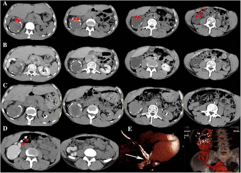
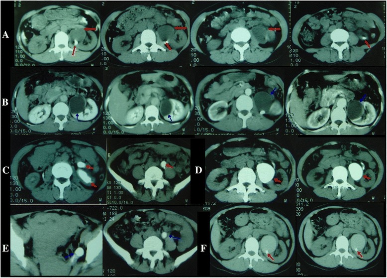
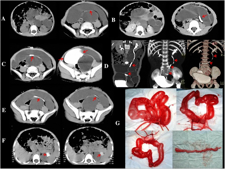
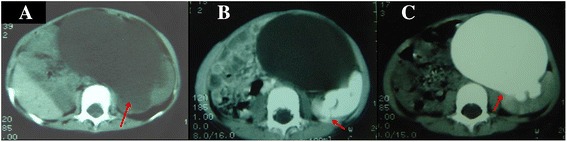
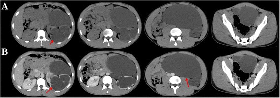
Background: Duplex kidney is a common anomaly that is frequently associated with multiple complications. Typical computed tomography urography (CTU) includes four phases (unenhanced, arterial, parenchymal and excretory) and has been suggested to considerably aid in the duplex kidney diagnosi. Unfortunately, regarding duplex kidney with prolonged dilatation, the affected parenchyma and tortuous ureters demonstrate a lack of or delayed excretory opacification. We used prolonged-delay CTU, which consists of another prolonged-delay phase (1- to 72-h delay; mean delay: 24 h) to opacify the duplicated ureters and affected parenchyma.
Methods: Seventeen patients (9 males and 8 females; age range: 2.5-56 y; mean age: 40.4 y) with duplex kidney were included in this study. Unenhanced scans did not find typical characteristics of duplex kidney, except for irregular perirenal morphology. Duplex kidney could not be confirmed on typical four-phase CTU, whereas it could be easily diagnosed in axial and CT-3D reconstruction using prolonged CTU (prolonged-delay phase).
Results: Between January 2005 and October 2010, in this review board-approved study (with waived informed consent), 17 patients (9 males and 8 females; age range: 2.5 ~ 56 y; mean age: 40.4 y) with suspicious duplex kidney underwent prolonged CTU to opacify the duplicated ureters and confirm the diagnosis.
Conclusion: Our results suggest the validity of prolonged CTU to aid in the evaluation of the function of the affected parenchyma and in the demonstration of urinary tract malformations.
Keywords: Duplex kidney; Duplicated ureters; Multi-slice spiral CT urography; Prolonged-delay contrast enhancement.
Figures
Similar articles
-
Excretory phase CT urography for opacification of the urinary collecting system.AJR Am J Roentgenol. 1998 May;170(5):1261-7. doi: 10.2214/ajr.170.5.9574598. AJR Am J Roentgenol. 1998. PMID: 9574598
-
Multi-detector CT urography: effect of oral hydration and contrast medium volume on renal parenchymal enhancement and urinary tract opacification--a quantitative and qualitative analysis.Eur Radiol. 2010 Sep;20(9):2146-52. doi: 10.1007/s00330-010-1785-8. Epub 2010 Apr 10. Eur Radiol. 2010. PMID: 20383638
-
CT evaluation of the upper urinary tract in adults younger than 50 years with asymptomatic microscopic hematuria: is IV contrast enhancement needed?AJR Am J Roentgenol. 2014 Sep;203(3):615-9. doi: 10.2214/AJR.13.11891. AJR Am J Roentgenol. 2014. PMID: 25148165
-
Computed tomographic urography update: an evolving urinary tract imaging modality.Semin Ultrasound CT MR. 2009 Aug;30(4):233-45. doi: 10.1053/j.sult.2009.03.005. Semin Ultrasound CT MR. 2009. PMID: 19711637 Review.
-
Tumors of Renal Collecting Systems, Renal Pelvis, and Ureters: Role of MR Imaging and MR Urography Versus Computed Tomography Urography.Magn Reson Imaging Clin N Am. 2019 Feb;27(1):15-32. doi: 10.1016/j.mric.2018.09.002. Magn Reson Imaging Clin N Am. 2019. PMID: 30466909 Review.
Cited by
-
First use and evaluation of a novel 6.3 Fr disposable flexible ureteroscope for stone management in duplex kidney: a case report.Transl Androl Urol. 2024 Nov 30;13(11):2644-2650. doi: 10.21037/tau-24-315. Epub 2024 Nov 27. Transl Androl Urol. 2024. PMID: 39698564 Free PMC article.
-
Extravesical Ectopic Ureteral Calculus Obstruction in a Completely Duplicated Collecting System.J Endourol Case Rep. 2020 Mar 11;6(1):42-44. doi: 10.1089/cren.2019.0108. eCollection 2020. J Endourol Case Rep. 2020. PMID: 32775673 Free PMC article.
-
Multi-Slice Spiral Computed Tomography Image Features under Hybrid Iterative Reconstruction Algorithm in Staging Diagnosis of Bladder Cancer.J Healthc Eng. 2021 Oct 27;2021:7733654. doi: 10.1155/2021/7733654. eCollection 2021. J Healthc Eng. 2021. Retraction in: J Healthc Eng. 2023 Oct 11;2023:9791241. doi: 10.1155/2023/9791241. PMID: 34745510 Free PMC article. Retracted.
-
CTU findings of duplex kidney in kidney: A rare duplicated renal malformation.Open Med (Wars). 2021 Apr 19;16(1):651-654. doi: 10.1515/med-2021-0271. eCollection 2021. Open Med (Wars). 2021. PMID: 33977147 Free PMC article.
-
Obstruction of bifid ureter by two calculi: A case report.Medicine (Baltimore). 2018 Jul;97(30):e11474. doi: 10.1097/MD.0000000000011474. Medicine (Baltimore). 2018. PMID: 30045270 Free PMC article.
References
MeSH terms
LinkOut - more resources
Full Text Sources
Other Literature Sources
Medical
