

Speckle-Tracking Echocardiography in Dogs With Patent Ductus Arteriosus: Effect of Percutaneous Closure on Cardiac Mechanics
- PMID: 27177625
- PMCID: PMC4913567
- DOI: 10.1111/jvim.13919
Speckle-Tracking Echocardiography in Dogs With Patent Ductus Arteriosus: Effect of Percutaneous Closure on Cardiac Mechanics
Abstract
Background: Patent ductus arteriosus (PDA) is 1 of the most common congenital heart defects in dogs and percutaneous closure is effective in achieving ductal closure; PDA closure is associated with abrupt hemodynamic changes.
Hypothesis: A marked decrease in standard parameters of systolic function as assessed by M- or B-mode echocardiography after PDA closure was identified in previous studies. Speckle tracking echocardiography can provide further insight into the effect of PDA closure on cardiac mechanics in dogs affected by PDA.
Animals: Twenty-five client-owned dogs with PDA.
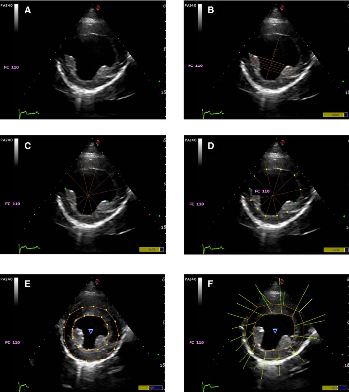
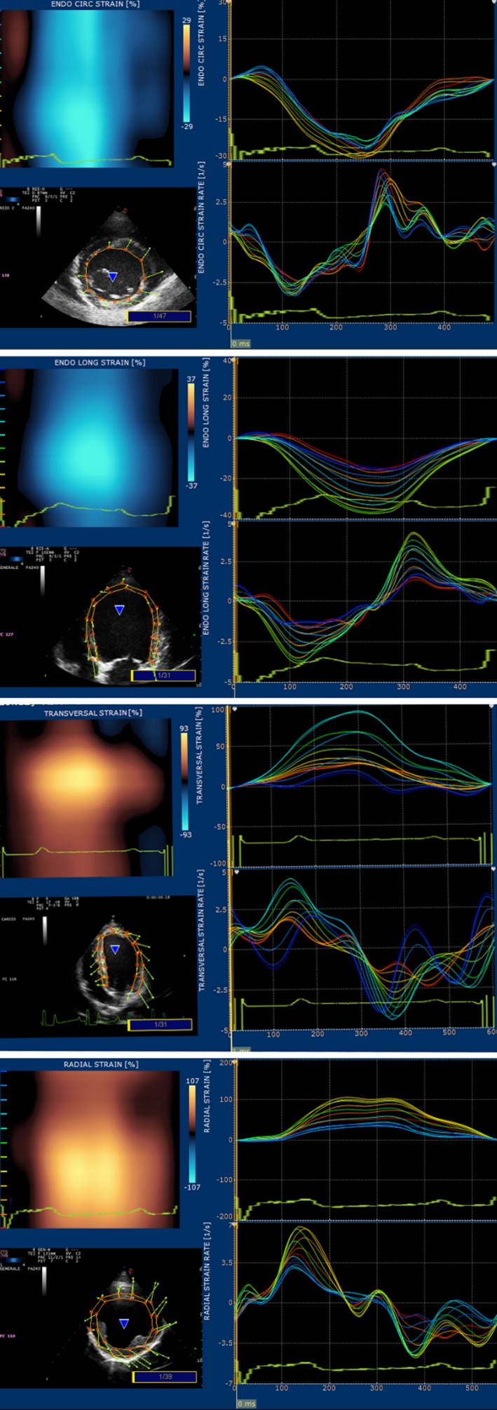
Methods: Prospective study. Dogs were recruited over a 2-year period. Complete echocardiographic evaluation was performed before and 24 hours after PDA closure, including standard (end-diastolic volumes indexed to body surface area in B- and M-mode [EDVIB /M ], end-systolic volumes indexed to body surface area in B- and M-mode [ESVIB /M ], allometric scaling in diastole [AlloD] and systole [AlloS], pulmonary flow to systemic flow [Qs/Qp], ejection fraction [EF], and fractional shortening [FS]), and advanced speckle-tracking echocardiography (STE): global longitudinal, radial, circumferential and transverse strain (S), and strain rate (SR).
Results: Patent ductus arteriosus closure was associated with statistically significant decreases in EDVIM /B and ESVIM /B , AlloD and AlloS, SI, EF, and FS. A statistically significant decrease in the absolute values of radial, transverse, and circumferential S and SR was observed, whereas longitudinal S and SR did not change significantly.
Conclusion and clinical importance: Patent ductus arteriosus closure by percutaneous approach is associated with marked decreases of conventional echocardiographic parameters as a result of the changes in loading conditions, but no evidence of systolic dysfunction was identified by means of STE, as none of the S and SR values were below reference ranges. In the short term, contractility is enhanced in the long axis (long S/SR values were not statistically different before and after closure) and decreases to normal values in short axis (circumferential, radial, and transversal S/SR decreased to normal reference range).
Keywords: Congenital heart disease; Dogs; Strain; Strain rate.
Copyright © 2016 The Authors. Journal of Veterinary Internal Medicine published by Wiley Periodicals, Inc. on behalf of the American College of Veterinary Internal Medicine.
Figures
Similar articles
-
Evaluation of left ventricular dimension and systolic function by standard transthoracic echocardiography before and 24-hours after percutaneous closure of patent ductus arteriosus in 120 dogs.PLoS One. 2019 Oct 9;14(10):e0223676. doi: 10.1371/journal.pone.0223676. eCollection 2019. PLoS One. 2019. PMID: 31596900 Free PMC article.
-
Echocardiographic Assessment of Cardiac Function by Conventional and Speckle-Tracking Echocardiography in Dogs with Patent Ductus Arteriosus.J Vet Intern Med. 2016 May;30(3):706-13. doi: 10.1111/jvim.13938. J Vet Intern Med. 2016. PMID: 27177624 Free PMC article.
-
Echocardiographic evaluation of myocardial changes observed after closure of patent ductus arteriosus in dogs.J Vet Intern Med. 2015 Jan;29(1):126-31. doi: 10.1111/jvim.12517. Epub 2015 Jan 16. J Vet Intern Med. 2015. PMID: 25594430 Free PMC article.
-
Echocardiography for patent ductus arteriosus including closure in adults.Echocardiography. 2015 Jan;32 Suppl 2:S125-39. doi: 10.1111/echo.12457. Epub 2014 May 29. Echocardiography. 2015. PMID: 24888537 Review.
-
Review of left-to-right shunting patent ductus arteriosus and short term outcome in 98 dogs.J Small Anim Pract. 2002 Sep;43(9):395-400. doi: 10.1111/j.1748-5827.2002.tb00090.x. J Small Anim Pract. 2002. PMID: 12238504 Review.
Cited by
-
Use of 2-dimensional speckle-tracking echocardiography to assess left ventricular systolic function in dogs with systemic inflammatory response syndrome.J Vet Intern Med. 2019 Mar;33(2):423-431. doi: 10.1111/jvim.15438. Epub 2019 Feb 17. J Vet Intern Med. 2019. PMID: 30773683 Free PMC article.
-
Speckle tracking echocardiography in cats with preclinical hypertrophic cardiomyopathy.J Vet Intern Med. 2019 May;33(3):1232-1241. doi: 10.1111/jvim.15495. Epub 2019 Apr 16. J Vet Intern Med. 2019. PMID: 30993757 Free PMC article.
-
Reference Intervals for Conventional Transthoracic Echocardiography and Two-Dimensional Speckle Tracking Echocardiography-Derived Strain Values in the Dutch Sheepdog ('Schapendoes').Animals (Basel). 2025 May 23;15(11):1524. doi: 10.3390/ani15111524. Animals (Basel). 2025. PMID: 40508989 Free PMC article.
-
A Retrospective Cohort Evaluation of Left Ventricular Remodeling, Perioperative Complications and Outcome in Medium and Large Size Dogs with Patent Ductus Arteriosus after Percutaneous Closure.Vet Sci. 2023 Nov 24;10(12):669. doi: 10.3390/vetsci10120669. Vet Sci. 2023. PMID: 38133219 Free PMC article.
-
Evaluation of left ventricular dimension and systolic function by standard transthoracic echocardiography before and 24-hours after percutaneous closure of patent ductus arteriosus in 120 dogs.PLoS One. 2019 Oct 9;14(10):e0223676. doi: 10.1371/journal.pone.0223676. eCollection 2019. PLoS One. 2019. PMID: 31596900 Free PMC article.
References
-
- Clyman RI. Mechanisms regulating the ductus arteriosus. Biol Neonate 2006;89:330–335. - PubMed
-
- Buchanan JW. Causes and prevalence of cardiovascular disease In: Kirk RW, Bonagura JD, eds. Kirk's Current Veterinary Therapy XI. Philadelphia, PA: WB Saunders, 1992:647–655.
-
- Buchanan JW. Prevalence of cardiovascular disorders In: Fox R, Sisson D, Moïse NS, eds. Textbook of Canine and Feline Cardiology, 2nd ed Philadelphia: Saunders; 1999:457–463.
-
- Tidholm A. Retrospective study of congenital heart defects in 151 dogs. J Small Anim Pract 1997;38:94–98. - PubMed
-
- Oliveira P, Domenech O, Bussadori C, et al. Retrospective review of congenital heart disease in 976 dogs. J Vet Int Med 2011;25:477–483. - PubMed
MeSH terms
LinkOut - more resources
Full Text Sources
Other Literature Sources
Research Materials
