. 2016:1441:151-65.
doi: 10.1007/978-1-4939-3684-7_13.

The Planar Lipid Bilayer System Serves as a Reductionist Approach for Studying NK Cell Immunological Synapses and Their Functions

Affiliations

The Planar Lipid Bilayer System Serves as a Reductionist Approach for Studying NK Cell Immunological Synapses and Their Functions

Grant Bertolet et al. Methods Mol Biol. 2016.

Abstract

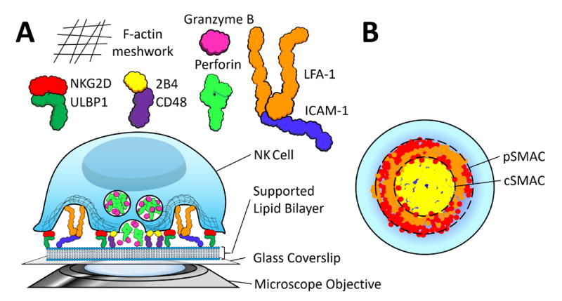
The immunological synapse (IS) is the junction between an immune cell (e.g., a T or NK cell) and another cell (e.g., an antigen-presenting cell (APC), or a tumor cell). The formation of the IS is crucial for cell-mediated immunity, and as such, an understanding of both the composition of the IS and the factors that drive its formation are essential for understanding how and when NK cells eliminate susceptible target cells. The supported lipid bilayer (SLB) system is a highly effective tool for directly studying the IS. SLBs confer three main advantages: (1) they allow for synapse formation on a level horizontal surface, allowing for direct visualization of the IS under high resolution imaging systems, (2) they mimic the surface of a target cell by providing a fluid mosaic into which surface proteins can be embedded while permitting free motion in two dimensions, which is important for studying the dynamics of synapse formation, and (3) they allow investigators to determine the exact composition of the bilayer, thus in turn allowing them to answer very specific questions about the IS. It is our hope that this chapter will furnish readers with an awareness of the applications of the SLB system for studying the IS in NK cells, and also of a basic knowledge of how to use this system for themselves.

Keywords: Confocal microscopy; IS; Immune synapse; Immunological synapse; Immunosynapse; NK; Natural killer; SLB; Supported lipid bilayer.


Figures

Figure 1
Figure 1
Cartoon illustrating a model of the NK cell synapse formed on the supported lipid bilayer (SLB). Ligands for various NK cell surface proteins are anchored in the SLB, which itself rests on a flat rigid glass surface. NK cells are added atop this bilayer, which they recognize much as they would the surface of a target cell, and an immunological synapses (IS) forms between the surface proteins on the NK cell and the ligands embedded in the SLB. The planar orientation of the bilayer allows for easy and direct imaging of the resulting IS. A] The particular synapse shown here is a model interpreted from the findings of Liu et al (10) and Zheng et al (13). On a lipid bilayer containing 1) ULBP1 (green), 2) CD48 (green), and 3) ICAM-1 (blue), these proteins will be bound by 1) NKG2D (red), 2) 2B4 (yellow), and 3) LFA-1 (orange), respectively, causing a re-organization of the F-actin cytoskeleton and polarization of perforin-positive lytic granules to the synapse. These lytic granules will then be extruded from the cell through regions of F-actin hypodensities, like salt granules through the pores in a shaker. B] The ‘head-on’ view of the IS provided by the SLB system allows for the visualization of the IS’ characteristic ‘bull’s-eye’ structure, which in this case consists of centralized aggregated 2B4 surrounded by an outer ring of LFA-1 and NKG2D. These regions are known as the cSMAC and pSMAC, respectively.
Figure 2
Figure 2
Overview of the principle behind determining the seeding density of a given protein on the SLB. Silica beads of known diameter are coated with the biotinylated phospholipids, followed by streptavidin, and then with biotinylated, fluorescently-labeled protein of various concentrations. These coated beads are then assayed by flow cytometry, and their fluorescent values are plotted against a standard curve established from a series of beads labeled with known numbers of the same fluorophore. From this, the number of molecules of equivalent soluble fluorochromes (MESF) can be correlated with the protein concentration, and dividing by the number of fluorophores per unit protein, the average density of protein coating each bead can be derived. From there, one simply needs to compute the surface area of the bead from its diameter to determine the seeding density of protein per square micrometer of the bilayer as a function of concentration.
Figure 3
Figure 3
How to calculate the MESF standard curve. A] Run a tube containing equal parts of each tube in the standard MESF series on a flow cytometer, then gate on the single beads in an SSC vs FSC dot plot. B] Plot the singlet population on a unidimensional histogram with the fluorescent channel on the X-axis, and draw a gate spanning the width of each peak at its half-maximal intensity. Record the arithmetic mean for each gate. C] Subtract the arithmetic mean for each peak from the blank value, then plot these against the MESF values (which can be found from the manufacturer). From this, a linear relationship between MESF and fluorescence can be obtained, which can then be used to calculate the MESF of your protein-labeled lipid-coated beads.
Figure 4
Figure 4
Overview of the process of constructing the SLB. The first step of the process is to add biotinylated lipids to a pre-treated glass surface. Then, streptavidin is added to provide a substrate for biotinylated proteins (here generically represented by an antibody). These proteins are strongly anchored onto the bilayer by the biotin-streptavidin interaction, but remain laterally mobile. Finally, cells are added to the bilayer, where they form protein-dependent contacts and, subsequently, immune synapses.
Figure 5
Figure 5
Illustration of how to position a coverslip vertically in order to air-dry, following cleaning with Piranha solution and subsequent rinsing with distilled water.
Figure 6
Figure 6
Illustration of how to place the coverslip over the bottom of the flow chamber. With ungloved hands, grasp the edges of the coverslip between your thumb and index finger as pictured. Line up the near edge of the coverslip with the near edge of the chamber, them gently and evenly lay down the coverslip away from yourself until it lies flat against the chamber.
Figure 7
Figure 7
Illustration of how to wash one chamber of the flow cell. A 1 mL pipette is placed at one end, and wash buffer is slowly passed through as suction is applied to the other end.

References

    1. Grakoui A, Bromley SK, Sumen C, et al. The immunological synapse: a molecular machine controlling T cell activation. Science. 1999;285(5425):221–7. - PubMed
    1. McConnell HM, Watts TH, Weis RM, et al. Supported planar membranes in studies of cell-cell recognition in the immune system. Biochim Biophys Acta. 1986;864(1):95–106. - PubMed
    1. Liu D, Bryceson YT, Meckel T, et al. Integrin-dependent organization and bidirectional vesicular traffic at cytotoxic immune synapses. Immunity. 2009;31(1):99–109. - PMC - PubMed
    1. Liu D, Peterson ME, Long EO. The adaptor protein Crk controls activation and inhibition of natural killer cells. Immunity. 2012;36(4):600–11. - PMC - PubMed
    1. Abeyweera TP, Merino E, Huse M. Inhibitory signaling blocks activating receptor clustering and induces cytoskeletal retraction in natural killer cells. J Cell Biol. 2011;192(4):675–90. - PMC - PubMed

LinkOut - more resources