. 2016 May 13;9(1):45.
doi: 10.1186/s13045-016-0275-0.

Calreticulin-mutant proteins induce megakaryocytic signaling to transform hematopoietic cells and undergo accelerated degradation and Golgi-mediated secretion

Affiliations

Calreticulin-mutant proteins induce megakaryocytic signaling to transform hematopoietic cells and undergo accelerated degradation and Golgi-mediated secretion

Lijuan Han et al. J Hematol Oncol. .

Abstract

Background: Somatic calreticulin (CALR), Janus kinase 2 (JAK2), and thrombopoietin receptor (MPL) mutations essentially show mutual exclusion in myeloproliferative neoplasms (MPN), suggesting that they activate common oncogenic pathways. Recent data have shown that MPL function is essential for CALR mutant-driven MPN. However, the exact role and the mechanisms of action of CALR mutants have not been fully elucidated.

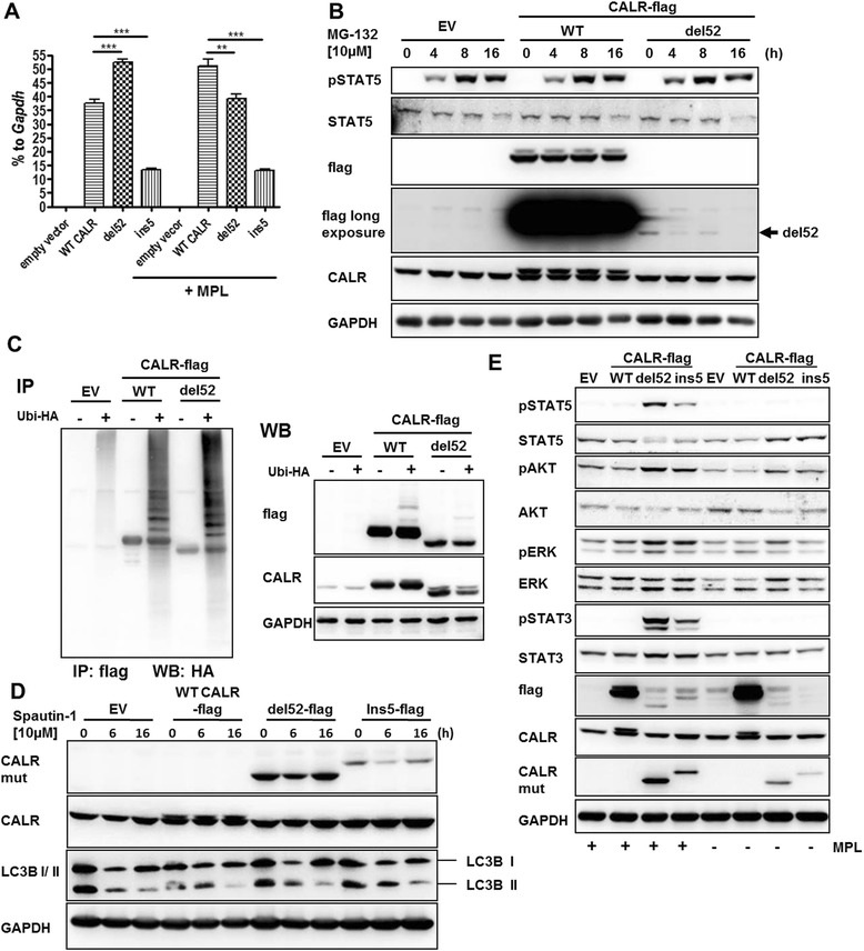
Methods: The murine myeloid cell line 32D and human HL60 cells overexpressing the most frequent CALR type 1 and type 2 frameshift mutants were generated to analyze the first steps of cellular transformation, in the presence and absence of MPL expression. Furthermore, mutant CALR protein stability and secretion were examined using brefeldin A, MG132, spautin-1, and tunicamycin treatment.

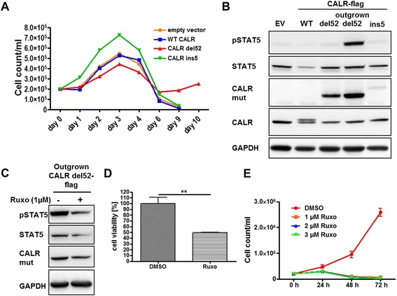
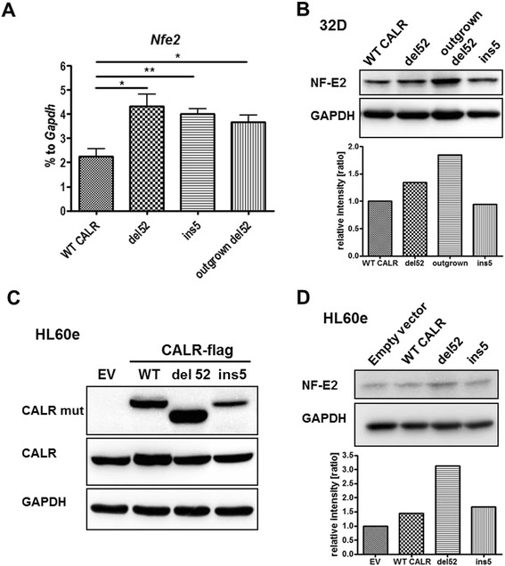
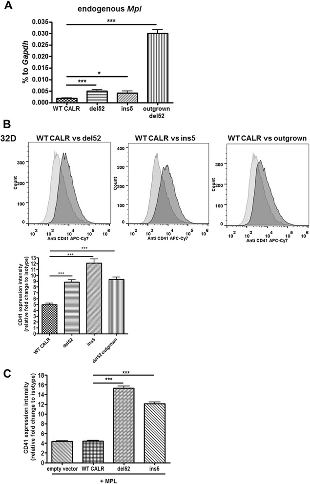
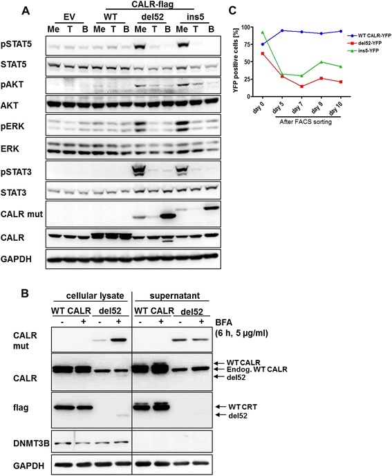
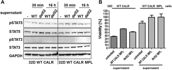
Results: The present study demonstrates that the expression of endogenous Mpl, CD41, and the key megakaryocytic transcription factor NF-E2 is stimulated by type 1 and type 2 CALR mutants, even in the absence of exogenous MPL. Mutant CALR expressing 32D cells spontaneously acquired cytokine independence, and this was associated with increased Mpl mRNA expression, CD41, and NF-E2 protein as well as constitutive activation of downstream signaling and response to JAK inhibitor treatment. Exogenous expression of MPL led to constitutive activation of STAT3 and 5, ERK1/2, and AKT, cytokine-independent growth, and reduction of apoptosis similar to the effects seen in the spontaneously outgrown cells. We observed low CALR-mutant protein amounts in cellular lysates of stably transduced cells, and this was due to accelerated protein degradation that occurred independently from the ubiquitin-proteasome system as well as autophagy. CALR-mutant degradation was attenuated by MPL expression. Interestingly, we found high levels of mutated CALR and loss of downstream signaling after blockage of the secretory pathway and protein glycosylation.

Conclusions: These findings demonstrate the potency of CALR mutants to drive expression of megakaryocytic differentiation markers such as NF-E2 and CD41 as well as Mpl. Furthermore, CALR mutants undergo accelerated protein degradation that involves the secretory pathway and/or protein glycosylation.

Keywords: Calreticulin; Degradation; Frameshift mutants; MPL; MPN; Megakaryopoiesis; Myeloproliferative neoplasms; NF-E2; Protein secretion; del52.


Figures

Fig. 1
Fig. 1
Spontaneously outgrown 32D CALR del52 cells are ruxolitinib (Ruxo) sensitive. a We performed growth assays with stably transduced 32D cells expressing empty vector (EV), WT CALR, del52 or the ins5 mutants (2 × 105 cells/ml). Outgrowth of a CALR del52 expressing clone has been observed after 6 to 10 days. b 32D EV, CALR WT, CALR del52, CALR ins5, and outgrown del52 cells were starved for 16 h; lysates were prepared and subjected to SDS-PAGE and Western blotting. Indicated antibodies have been used for immunostaining. c Outgrown 32D CALR del52 cells were treated overnight with 1 μM Ruxo, and reduction of STAT5 phosphorylation could be detected in Western blotting. d In a MTT assay, outgrown 32D CALR del52 cells were treated with 1 μM Ruxo for 48 h, and cell viability was evaluated. The experiment was performed in triplicates. SD is indicated. **P < 0.01 e Outgrown 32D del52 cells were grown in WEHI-free medium (2 × 105 cells/ml) supplemented with 1, 2, and 3 μM Ruxo or DMSO as control for up to 72 h. The cells were counted every 24 h. The experiment was performed in triplicates. SD is indicated
Fig. 2
Fig. 2
32D cells expressing CALR mutants show increased mRNA and protein levels of the megakaryocytic transcription factor NF-E2. a Detection of Nfe2 mRNA expression by RT-qPCR in the indicated 32D cells. The experiments were performed in triplicates. SD is indicated. *P < 0.05, **P < 0.01, ***P < 0.001. b Lysates were prepared of 32D cells expressing WT CALR, del52, and ins5 mutant as well as of outgrown 32D del52 cells, and NF-E2 protein was detected in Western blotting. In b and d, GAPDH served as loading control and was used for the calculation of NF-E2 expression ratios. c HL60e EV (empty vector), CALR WT, CALR del52 and CALR ins5 cells were used to prepare lysates, and SDS-PAGE and Western blotting were performed. Indicated antibodies have been used for immunostaining. CALR mut antibody showed unspecific binding to ectopic WT CALR. d HL60e cells expressing empty vector (EV), WT CALR, del52, or ins5 were analyzed for NF-E2 expression, and expression ratios were calculated as in b
Fig. 3
Fig. 3
Endogenous Mpl as well as CD41 was upregulated by CALR-mutant expression. a RT-qPCR was used to detect Mpl mRNA amounts after RNA isolation of the indicated 32D cells followed by cDNA synthesis. Expression is depicted in percentage to Gapdh. Measurements were done in triplicates. SD is indicated. *P < 0.05 b FACS analysis of membrane-localized CD41 in the indicated 32D cell lines. Relative expression intensity was calculated using the mean fluorescent intensity (MFI) of triplicates. SD is indicated. ***P < 0.001 c As in b, the amount of CD41 was evaluated in 32D MPL cells expressing empty vector, WT CALR, del52, or ins5 using FACS. The experiment was performed in triplicates and the SD is indicated. ***P < 0.001
Fig. 4
Fig. 4
Degradation of instable CALR mutants is proteasome-independent and mutants get stabilized by MPL expression. a The expression of indicated CALR-flag constructs in 32D cells +/− ectopic MPL was confirmed by RT-qPCR. The expression is depicted as percentage to Gapdh. Experiments were performed in triplicates. Mean and SD are indicated. **P < 0.01, ***P < 0.001. b 32D empty vector (EV), WT CALR-positive, or del52-positive cells were treated with 10 μM MG132 for indicated periods of time. Afterwards, cells were starved in WEHI-free medium for 4 h and lysates were prepared for SDS-PAGE and Western blotting. Indicated antibodies were used for immunodetection to show protein stability. c HEK293T cells were transfected with HA-tagged ubiquitin and the indicated CALR-flag constructs. After 24 h, 10 μM MG132 was added for 20 h and protein lysates were prepared. Immunoprecipitation (IP) was performed with flag antibody followed by SDS-PAGE and Western blotting. In addition, whole cell lysates were used for Western blotting. d Autophagosomal inhibition was performed in 32D EV, WT CALR, del52, and ins5 expressing cells. The 32D cell lines were treated with the inhibitor spautin-1 for the indicated time. Lysates were prepared, and SDS-PAGE and Western blotting were performed. Antibodies detecting mutated CALR (CALR mut), CALR, LC3I-II, and GAPDH were used for immunostaining. LC3I-II served as control for successful autophagosomal inhibition. e 32D cells expressing EV, WT CALR, del52, or ins5 mutant +/− MPL receptor were starved overnight. Lysates were prepared and subjected to SDS-PAGE and Western blotting. The PVDF membrane was subjected to the indicated antibodies
Fig. 5
Fig. 5
Glycosylation of MPL is essential for the activatory mechanism of CALR mutants, which are strongly secreted. a Empty vector (EV), WT CALR, del52, and ins5 expressing 32D MPL cells were treated with tunicamycin (T; 10 μg/ml), brefeldin A (B; 5 μg/ml), or the solvent methanol (Me) as control. Lysate preparation, SDS-PAGE, and Western blotting were performed. Indicated antibodies were used to confirm downstream signaling and CALR expression. GAPDH staining served as loading control. b Concentrated supernatants (30 μg protein) of indicated 32D cells, which were treated for 6 h with 5 μg/ml BFA or left untreated, were applied to SDS-PAGE followed by Western blotting and compared to the appropriate cellular lysates (30 μg protein). c 32D MPL cells expressing C-terminally YFP-tagged WT CALR, del52, or ins5 were FACS sorted for equal YFP-intensities and YFP signal was monitored until 10 days after sorting. Strong decrease of CALR del52-flag-YFP and ins5-flag-YFP could be observed
Fig. 6
Fig. 6
Secreted CALR del52 protein shows no paracrine function in 32D MPL cells. a 32D WT CALR +/− MPL cells were cultured with the indicated supernatants (12 μg of protein) for 30 min or 16 h before preparation of lysates to analyze STAT5 and STAT3 phosphorylation in Western blotting. b MTT assays were performed to analyze viability of 32D WT CALR +/− MPL cells cultured in FBS and WEHI-free medium for 48 h treated with the indicated supernatants (3.5 μg/100 μl). Relative viability in percent was calculated by determination of the values gained by the 32D MPL WT CALR cells + del52 supernatant approach as 100 %

References

    1. Nangalia J, Massie CE, Baxter EJ, Nice FL, Gundem G, Wedge DC, et al. Somatic CALR mutations in myeloproliferative neoplasms with nonmutated JAK2. N Engl J Med. 2013;369:2391–405. doi: 10.1056/NEJMoa1312542. - DOI - PMC - PubMed
    1. Klampfl T, Gisslinger H, Harutyunyan AS, Nivarthi H, Rumi E, Milosevic JD, et al. Somatic mutations of calreticulin in myeloproliferative neoplasms. N Engl J Med. 2013;369:2379–90. doi: 10.1056/NEJMoa1311347. - DOI - PubMed
    1. Tefferi A, Barbui T. Polycythemia vera and essential thrombocythemia: 2015 update on diagnosis, risk-stratification and management. Am J Hematol. 2015;90:162–73. doi: 10.1002/ajh.23895. - DOI - PubMed
    1. Beer P. The pathogenesis of essential thrombocythemia. Curr Opin Hematol. 2011;18:323–9. doi: 10.1097/MOH.0b013e3283497f54. - DOI - PubMed
    1. Broséus J, Park J-H, Carillo S, Hermouet S, Girodon F. Presence of calreticulin mutations in JAK2-negative polycythemia vera. Blood. 2014;124:3964–6. doi: 10.1182/blood-2014-06-583161. - DOI - PubMed

MeSH terms

LinkOut - more resources