

Enhanced incorporation of dietary DHA into lymph phospholipids by altering its molecular carrier
- PMID: 27178174
- PMCID: PMC4911702
- DOI: 10.1016/j.bbalip.2016.05.002
Enhanced incorporation of dietary DHA into lymph phospholipids by altering its molecular carrier
Abstract
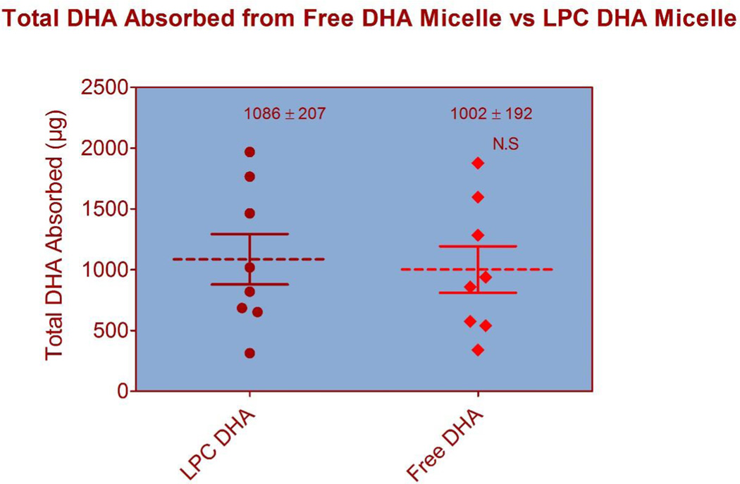
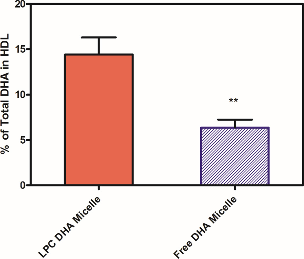
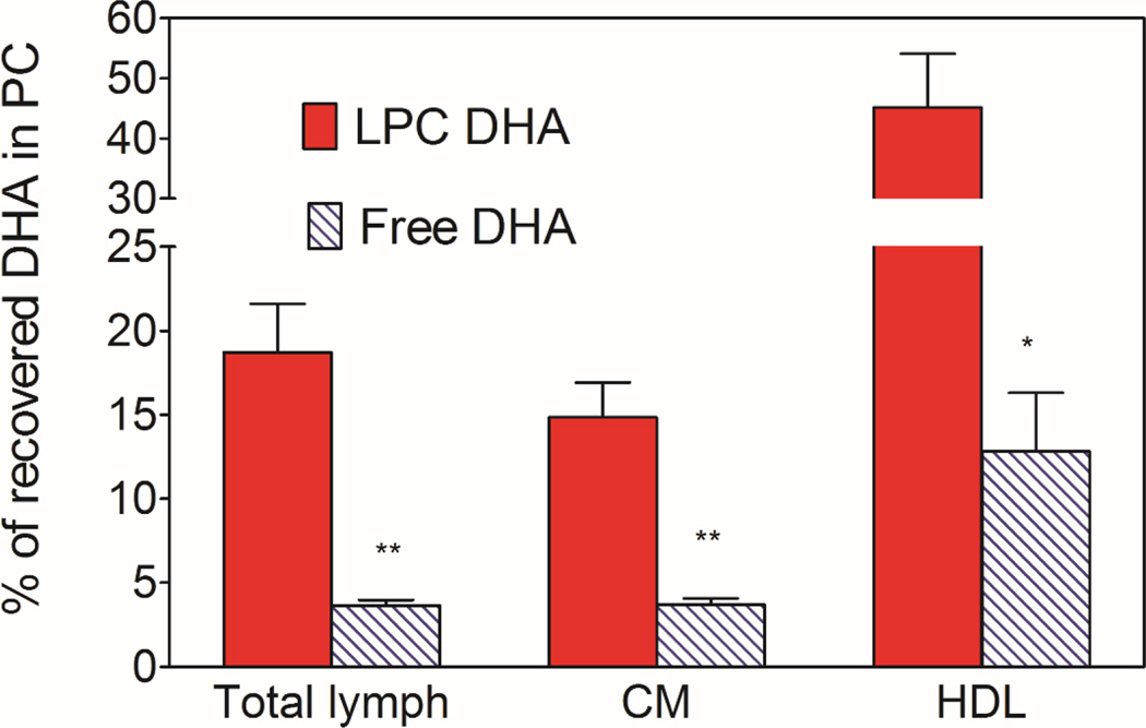
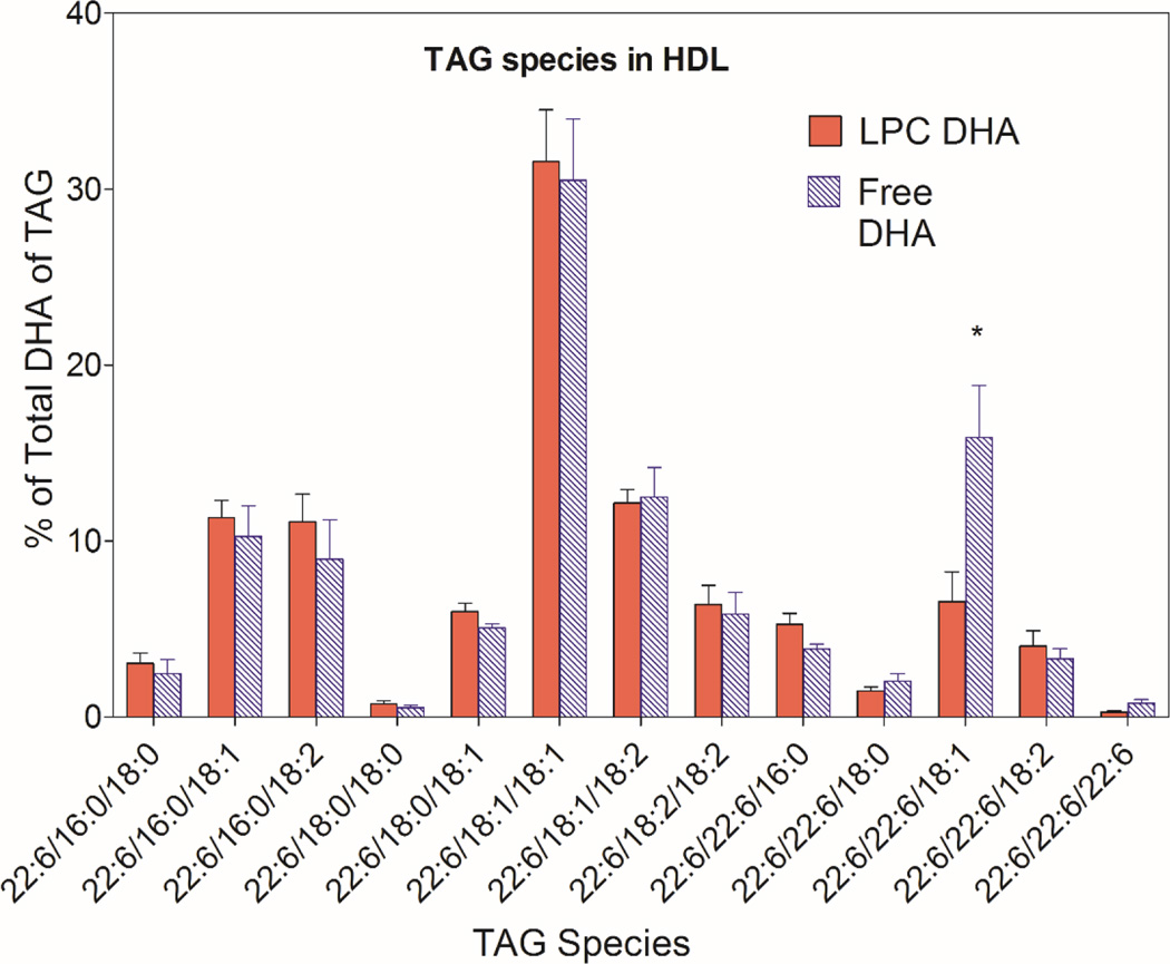
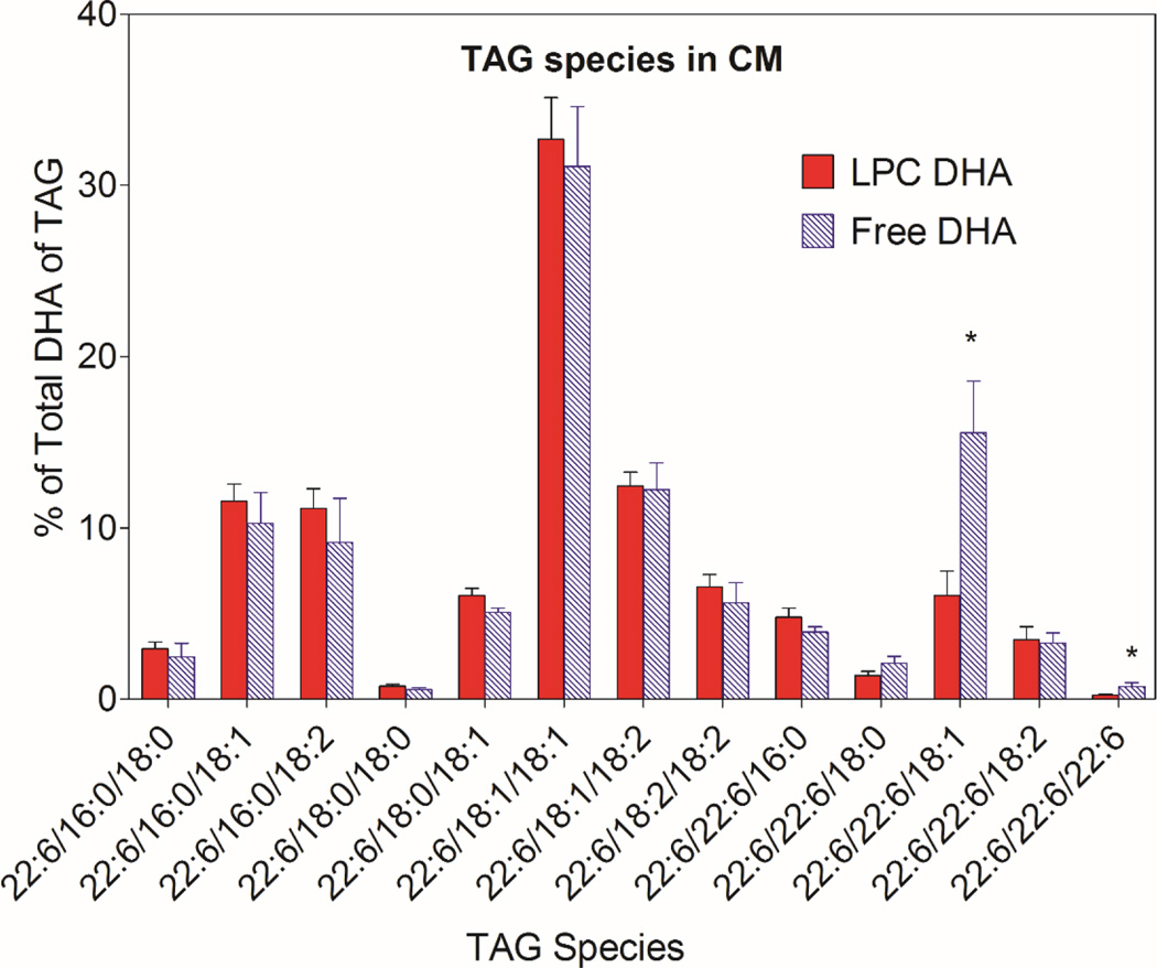
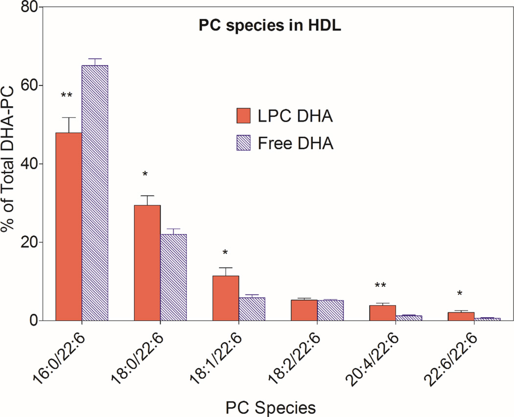
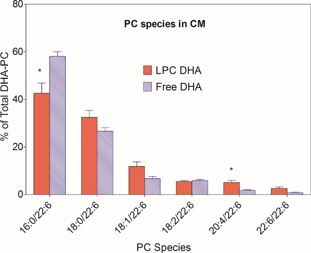
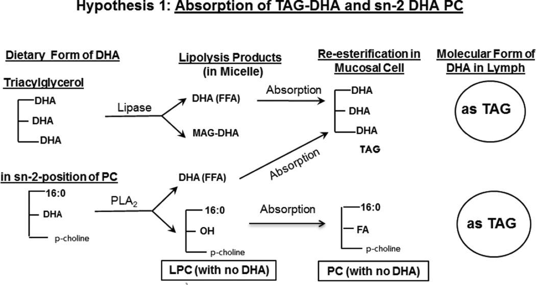
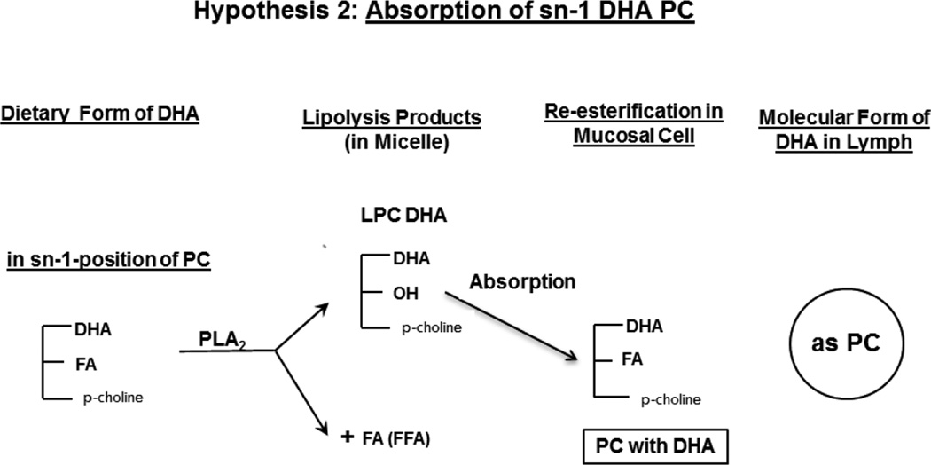
Several previous studies indicated that for optimal uptake by the brain, docosahexaenoic acid (DHA) should be present as phospholipid in the plasma. However most of dietary DHA is absorbed as triacylglycerol (TAG) because it is released as free fatty acid during digestion of either TAG-DHA (fish oil) or sn-2-DHA phospholipid (krill oil), and subsequently incorporated into TAG of chylomicrons. We tested the hypothesis that the absorption of DHA as phospholipid can be increased if it is present in the sn-1 position of dietary phospholipid or in lysophosphatidylcholine (LPC), because it would escape the hydrolysis by pancreatic phospholipase A2. We infused micelle containing the DHA either as LPC or as free acid, into the duodenum of lymph cannulated rats, and analyzed the chylomicrons and HDL of the lymph for the DHA-containing lipids. The results show that while the total amount of DHA absorbed was comparable from the two types of micelle, the percentage of DHA recovered in lymph phospholipids was 5 times greater with LPC-DHA, compared to free DHA. Furthermore, the amount of DHA recovered in lymph HDL was increased by 2-fold when LPC-DHA micelle was infused. These results could potentially lead to a novel strategy to increase brain DHA levels through the diet.
Keywords: Chylomicrons/HDL; Fish oil/DHA; Lymph; Lysophosphatidylcholine; Micelles; Phospholipids/absorption.
Published by Elsevier B.V.
Figures

Similar articles
-
Roe-derived phospholipid administration enhances lymphatic docosahexaenoic acid-containing phospholipid absorption in unanesthetized rats.Prostaglandins Leukot Essent Fatty Acids. 2018 Dec;139:40-48. doi: 10.1016/j.plefa.2017.06.011. Epub 2017 Jun 28. Prostaglandins Leukot Essent Fatty Acids. 2018. PMID: 28684067
-
Gastric digestion modifies absorption of butterfat into lymph chylomicrons in rats.J Nutr. 1998 Dec;128(12):2403-10. doi: 10.1093/jn/128.12.2403. J Nutr. 1998. PMID: 9868188
-
Enrichment of brain docosahexaenoic acid (DHA) is highly dependent upon the molecular carrier of dietary DHA: lysophosphatidylcholine is more efficient than either phosphatidylcholine or triacylglycerol.J Nutr Biochem. 2019 Dec;74:108231. doi: 10.1016/j.jnutbio.2019.108231. Epub 2019 Aug 31. J Nutr Biochem. 2019. PMID: 31665653 Free PMC article.
-
Marine Fish-Derived Lysophosphatidylcholine: Properties, Extraction, Quantification, and Brain Health Application.Molecules. 2023 Mar 30;28(7):3088. doi: 10.3390/molecules28073088. Molecules. 2023. PMID: 37049852 Free PMC article. Review.
-
Lysophosphatidylcholine as a preferred carrier form of docosahexaenoic acid to the brain.J Mol Neurosci. 2001 Apr-Jun;16(2-3):201-4; discussion 215-21. doi: 10.1385/JMN:16:2-3:201. J Mol Neurosci. 2001. PMID: 11478375 Review.
Cited by
-
Overconsumption of Omega-6 Polyunsaturated Fatty Acids (PUFAs) versus Deficiency of Omega-3 PUFAs in Modern-Day Diets: The Disturbing Factor for Their "Balanced Antagonistic Metabolic Functions" in the Human Body.J Lipids. 2021 Mar 17;2021:8848161. doi: 10.1155/2021/8848161. eCollection 2021. J Lipids. 2021. PMID: 33815845 Free PMC article. Review.
-
Role of Omega-3 and Omega-6 on Cardiovascular Risk Factors: Importance of Dietary Sources and Lipid Structure.Arq Bras Cardiol. 2023 Nov;120(11):e20230753. doi: 10.36660/abc.20230753. Arq Bras Cardiol. 2023. PMID: 38126490 Free PMC article. English, Portuguese. No abstract available.
-
Dietary lysophosphatidylcholine-EPA enriches both EPA and DHA in the brain: potential treatment for depression.J Lipid Res. 2019 Mar;60(3):566-578. doi: 10.1194/jlr.M090464. Epub 2018 Dec 10. J Lipid Res. 2019. PMID: 30530735 Free PMC article.
-
Role of phosphatidylcholine-DHA in preventing APOE4-associated Alzheimer's disease.FASEB J. 2019 Feb;33(2):1554-1564. doi: 10.1096/fj.201801412R. Epub 2018 Oct 5. FASEB J. 2019. PMID: 30289748 Free PMC article. Review.
-
Novel approach to enhancing brain DHA uptake: the role of nannochloropsis microalgae extract.Front Nutr. 2025 Jul 9;12:1573310. doi: 10.3389/fnut.2025.1573310. eCollection 2025. Front Nutr. 2025. PMID: 40704302 Free PMC article.
References
-
- Alessandri JM, Guesnet P, Vancassel S FAU - Astorg P, Astorg PF, Denis IF, Langelier BF, Aid S FAU - Poumes-Ballihaut C, Poumes-Ballihaut CF, Champeil-Potokar GF, Lavialle M. Polyunsaturated fatty acids in the central nervous system: evolution of concepts and nutritional implications throughout life. Reprod. Nutr Dev. 2004;44:509–538. - PubMed
-
- Chen CT, MA DWL, Kim JH, Mount HTJ, Bazinet RP. The low density lipoprotein receptor is not necessary for maintaining mouse brain polyunsaturated fatty acid concentrations. J. Lipid Res. 2008;49:147–152. - PubMed
-
- Rahman T, Taha AY, Jun Song B, Orr SK, Liu Z, Chen CT, Bazinet RP. The very low density lipoprotein receptor is not necessary for maintaining brain polyunsaturated fatty acid concentrations. Prostaglandins, Leukotrienes and Essential Fatty Acids. 2010;82:141–145. - PubMed
-
- Thies F, Pillon C, Moliere P, Lagarde M, Lecerf J. Preferential incorporation of sn-2 lysoPC DHA over unesterified DHA in the young rat brain. Amer. J. Physiol-Regul. Integr. C. 1994;36:R1273–R1279. - PubMed
Publication types
MeSH terms
Substances
Grants and funding
LinkOut - more resources
Full Text Sources
Other Literature Sources
Medical
