. 2016 May 13;13(1):107.
doi: 10.1186/s12974-016-0571-1.

Interferon gamma protects neonatal neural stem/progenitor cells during measles virus infection of the brain

Affiliations

Interferon gamma protects neonatal neural stem/progenitor cells during measles virus infection of the brain

Kristen N Fantetti et al. J Neuroinflammation. .

Abstract

Background: In the developing brain, self-renewing neural stem/progenitor cells (NSPC) give rise to neuronal and glial lineages. NSPC survival and differentiation can be altered by neurotropic viruses and by the anti-viral immune response. Several neurotropic viruses specifically target and infect NSPCs, in addition to inducing neuronal loss, which makes it difficult to distinguish between effects on NSPCs that are due to direct viral infection or due to the anti-viral immune response.

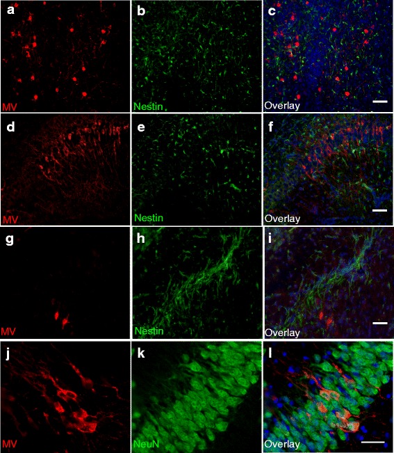
Methods: We have investigated the impact of anti-viral immunity on NSPCs in measles virus (MV)-infected neonates. A neuron-restricted viral infection model was used, where NSPCs remain uninfected. Thus, an anti-viral immune response was induced without the confounding issue of NSPC infection. Two-transgenic mouse lines were used: CD46+ mice express the human isoform of CD46, the MV entry receptor, under the control of the neuron-specific enolase promoter; CD46+/IFNγ-KO mice lack the key anti-viral cytokine IFNγ. Multi-color flow cytometry and Western Blot analysis were used to quantify effects on NSPC, neuronal, and glial cell number, and quantify effects on IFNγ-mediated signaling and cell markers, respectively.

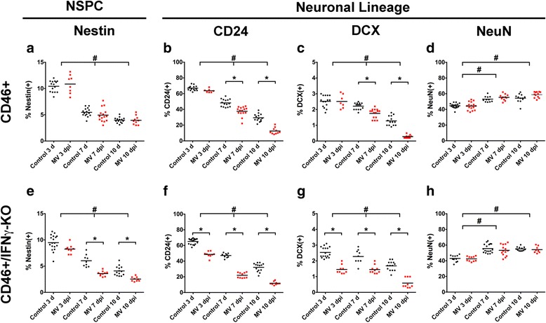
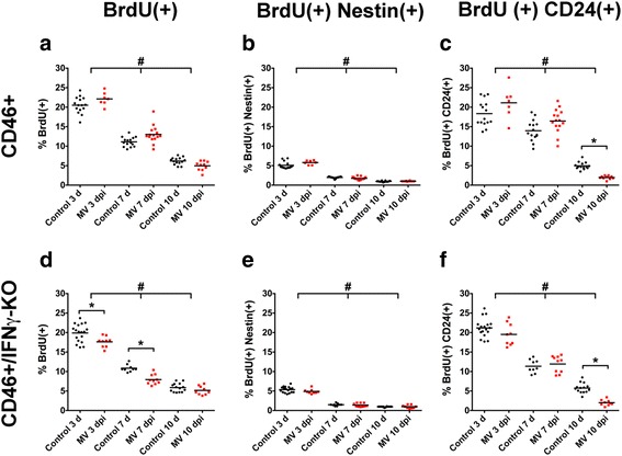
Results: Flow cytometric analysis revealed that NSPCs were reduced in CD46+/IFNγ-KO mice at 3, 7, and 10 days post-infection (dpi), but were unaffected in CD46+ mice. Early neurons showed the greatest cell loss at 7 dpi in both genotypes, with no effect on mature neurons and glial cells. Thus, IFNγ protected against NSPC loss, but did not protect young neurons. Western Blot analyses on hippocampal explants showed reduced nestin expression in the absence of IFNγ, and reduced doublecortin and βIII-tubulin in both genotypes. Phosphorylation of STAT1 and STAT2 occurred independently of IFNγ in the hippocampus, albeit with distinct regulation of activation.

Conclusions: This is the first study to demonstrate bystander effects of anti-viral immunity on NSPC function. Our results show IFNγ protects the NSPC population during a neonatal viral CNS infection. Significant loss of NSPCs in CD46+/IFNγ-KO neonates suggests that the adaptive immune response is detrimental to NSPCs in the absence of IFNγ. These results reveal the importance and contribution of the anti-viral immune response to neuropathology and may be relevant to other neuroinflammatory conditions.

Keywords: Anti-viral; Glia; Immune response; Interferon-γ; Measles virus; Neonate; Neural stem cell; Neurogenesis; STAT signaling.


Figures

Fig. 1
Fig. 1
MV infects neurons, but not NSPCs, in CD46+ mice. Sagittal brain sections from MV-infected CD46+ mice were collected at 10 days post-infection (dpi) and stained for MV (red) and nestin (green; ai) or MV (red) and NeuN (green; jl). All sections were stained with Hoechst (blue) to visualize nuclei. Representative images are shown from different brain regions, including the thalamus (ac), CA1 region of the hippocampus (df), fiber tracts of the medial forebrain bundle system (gi), and the dentate gyrus (jl). MV staining was restricted to NeuN+ cells (jl). Co-localization with nestin and MV was not observed in any brain region (ai). Panels ai: Magnification = ×20; scale bar = 50 μm. Panels jl: Magnification = ×40; scale bar = 25 μm
Fig. 2
Fig. 2
IFNγ preserves the NSPC population, but not early neurons. CD46+ (ad) and CD46+/IFNγ-KO (eh) control and MV-infected neonatal whole-brain isolates labeled for multipotent NSPCs (nestin: a, e) and cells specified to the neuronal lineage (CD24: b, f; DCX: c, g; NeuN: d, h), at 3, 7, and 10 dpi. The percentage of nestin + cells was significantly reduced at 7 and 10 dpi in MV-infected IFNγ-KO (e), but not MV-infected CD46+ mice (a). Significant CD24+ (b, f) and DCX+ (c, g) immature neuron cell loss occurred in both genotypes. Mature NeuN+ cell number was unaffected (d, h). *p < 0.01 significantly different uninfected versus MV-infected; #p < 0.05 significantly different between dpi
Fig. 3
Fig. 3
IFNγ does not alter glial differentiation. CD46+ (ad) and CD46+/IFNγ-KO (e-h) control and MV-infected neonatal whole-brain isolates were labeled for early glial precursors (A2B5: a, e), oligodendrocytes (O4: b, f) and astrocytes (GFAP: c, g). The average GFAP mean fluorescent intensity (MFI) was quantified and normalized to the MFI of the isotype control (d, h). There was no significant difference between control and MV-infected pups for the glial cell populations in either genotype. A significant decrease in %A2B5+ cells was observed in CD46+ (a), but not IFNγ-KO mice (d) mice from 3 to 10 dpi and a significant increase in O4+ oligodendrocytes was observed from 3 to 10 dpi in both genotypes (b, e). Age-dependent differences in GFAP expression also occurred (d, h). *p < 0.01 significantly different uninfected versus MV-infected; #p < 0.05
Fig. 4
Fig. 4
MV infection does not alter BrdU uptake by NSPCs. CD46+ (a-c) and CD46+/IFNγ-KO (d-f) pups were injected with BrdU 24 h before whole-brain isolates were processed and labeled for BrdU+ cells (a, d), or BrdU(+)Nestin(+) (b, e), and BrdU(+)CD24(+) (c, f) double-positive cells. Reduced BrdU uptake was observed in MV-infected CD46+/IFNγ-KO pups at 3 and 7 dpi (d), but not CD46+ mice (a). BrdU+ cells within the Nestin+ population showed no significant difference between control and MV-infected pups in both genotypes, but a significant reduction with age (b, e). The percentage of BrdU+ cells within the CD24+ cell population was reduced at 10 dpi (c, f), when severe illness and death occurs. *p < 0.01 significantly different uninfected versus MV-infected; #p < 0.05 significantly different between dpi
Fig. 5
Fig. 5
MV infection induces extensive apoptosis, but does not alter nestin localization. CD46+ and CD46+/IFNγ-KO brains were assayed by TUNEL (ac) or immunohistochemistry for nestin and NeuN at 7 dpi (dg). The average TUNEL+ cells per field (four fields per slice) were averaged from three different sagittal sections per mouse (c; n = 3 mice per condition). Significant increases in TUNEL+ cells were seen during infection in both genotypes, but no significant difference was observed between genotypes (c; *p < 0.01 significantly different uninfected versus MV-infected). Nestin (red) and NeuN (green) staining was observed in the hippocampus at 7 dpi in both CD46+ (d, e) and CD46+/IFNγ-KO (f, g) mice during infection (scale bar = 100 μm)
Fig. 6
Fig. 6
Impact of MV infection on expression of NSPC, neuronal, and glial markers in the hippocampus. Whole tissue lysates of hippocampal explants from CD46+ (ad) and CD46+/IFNγ-KO (eh) control and MV-infected mice were analyzed by Western Blots for Nestin (NSPC; ae), DCX (early neuron; b, f), βIII-tubulin (mature neuron; c, g) and GFAP (astrocyte; d, h) proteins. Integrated intensity values for each band, which are proportional to the sum of the fluorescent signal (measured in pixels) enclosed by the region of interest, are normalized to GAPDH as a protein loading control. Changes in nestin and DCX hippocampal protein expression correspond with changes in cell number observed for whole-brain cell isolates. *p < 0.01 significantly different uninfected versus MV-infected; #p < 0.05 significantly different between dpi
Fig. 7
Fig. 7
IFNγ does not reduce measles virus antigen in neonatal brain. a. Lysates of brain tissue from CD46+ and CD46+/IFNγ-KO neonates were analyzed by Western Blot for measles virus antigen. Control and MV-infected neonatal hippocampal lysates were collected at 3, 7, and 10 days post-infection (dpi) and probed with human polyclonal serum against MV and GAPDH as a loading control. b. Quantitation of MV levels normalized to GAPDH for each treatment condition in a. Statistical analysis was applied by two-way ANOVA (n = 4; *p < 0.01)
Fig. 8
Fig. 8
STAT1α and STAT1β isoforms in the hippocampus are phosphorylated in the absence of IFNγ during a viral infection. Lysates of hippocampal explants from CD46+ (ag) and CD46+/IFNγ-KO (hn) control and MV-infected mice were analyzed by western blot for phosphorylated (activated) STAT1 (b, c, i, j), and total STAT1 (d, e, k, l) proteins. GAPDH was used as a protein loading control. Representative blots are shown in a and b. Total levels of STAT1α (upper band; d, k) and STAT1β (lower band; e, l) were significantly increased in MV-infected hippocampal explants from CD46+ pups (d, e) at 7 and 10 dpi and in CD46+/IFNγ-KO pups (k, l) at 10 dpi. Phosphorylation of STAT1α (STAT1α-P; B, I) increased significantly in CD46+ explants at 7 and 10 dpi (b) and CD46+/IFNγ-KO explants at 10 dpi (I). Phosphorylation of STAT1β (c, j) was increased in CD46+ explants at 7 dpi only (c) and in CD46+/IFNγ-KO explants and 7 and 10 dpi (j). Protein ratios of STAT1α-P/STAT1α showed increased activation of phosphorylation of STAT1α-P at 10 dpi in CD46+ mice (f), but no activation in CD46+/IFNγ-KO mice (m). The protein ratios of STAT1β-P/STAT1β showed decreased activation of phosphorylation during infection in CD46+/IFNγ-KO mice (n), but not in CD46+ mice (g). Statistical analysis was applied by one-way ANOVA with multiple comparisons. (**p < 0.01, ***p < 0.001, ****p < 0.0001 significantly different uninfected versus MV-infected; n = 4)
Fig. 9
Fig. 9
STAT2, but not STAT3, is activated during MV infection in the neonatal hippocampus. Lysates of hippocampal explants from CD46+ (black bars; b, d, f, I, k, m) and CD46+/IFNγ-KO (grey bars; c, e, g, j, l, n) control and MV-infected mice were analyzed by western blot for phosphorylated (activated) STAT2 (STAT2-P; b, c), total STAT2 (d, e), STAT2-P/STAT2 (f, g), STAT3-P (i, j), total STAT3 (k, l), and STAT3-P/STAT3 (m, n). GAPDH was used as a protein loading control. Representative blots are shown for STAT2 (a) and STAT3 (h). STAT2-P levels were significantly increased in MV-infected hippocampal explants in CD46+ (b) and CD46+/IFNγ-KO (c) pups at 7 dpi, though STAT2 phosphorylation was sustained to 10 dpi in CD46+ explants. Protein ratios of STAT2-P/STAT2 showed significantly increased activation of phosphorylation in CD46+ mice at 3 and 7 dpi (f). STAT3 phosphorylation was not significantly induced under any condition (g, h, m, n). Levels of total STAT3 protein were increased with infection at 10 dpi in CD46+ explants (i), but not in CD46+/IFNγ-KO explants (j). Statistical analysis was applied by one-way ANOVA with multiple comparisons (*p < 0.05, ***p < 0.001, ****p < 0.0001 significantly different uninfected versus MV-infected; n = 4)

Similar articles

Cited by

References

    1. Schleede L, Bueter W, Baumgartner-Sigl S, Opladen T, Weigt-Usinger K, Stephan S, Smitka M, Leiz S, Kaiser O, Kraus V, et al. Pediatric herpes simplex virus encephalitis: a retrospective multicenter experience. J Child Neurol. 2013;28:321–31. doi: 10.1177/0883073812471428. - DOI - PubMed
    1. Bonthius DJ. Lymphocytic choriomeningitis virus: a prenatal and postnatal threat. Adv Pediatr. 2009;56:75–86. doi: 10.1016/j.yapd.2009.08.007. - DOI - PubMed
    1. Tardieu M, Le Chenadec J, Persoz A, Meyer L, Blanche S, Mayaux MJ. HIV-1-related encephalopathy in infants compared with children and adults. French Pediatric HIV Infection Study and the SEROCO Group. Neurology. 2000;54:1089–95. doi: 10.1212/WNL.54.5.1089. - DOI - PubMed
    1. Douvoyiannis M, Litman N, Goldman DL. Neurologic manifestations associated with parvovirus B19 infection. Clin Infect Dis. 2009;48:1713–23. doi: 10.1086/599042. - DOI - PubMed
    1. Allsopp TE, Scallan MF, Williams A, Fazakerley JK. Virus infection induces neuronal apoptosis: a comparison with trophic factor withdrawal. Cell Death Differ. 1998;5:50–9. doi: 10.1038/sj.cdd.4400298. - DOI - PubMed

MeSH terms