. 2016 May 17;44(5):1151-61.
doi: 10.1016/j.immuni.2016.04.009. Epub 2016 May 10.

Ubiquitination of the Transcription Factor IRF-3 Activates RIPA, the Apoptotic Pathway that Protects Mice from Viral Pathogenesis

Affiliations

Ubiquitination of the Transcription Factor IRF-3 Activates RIPA, the Apoptotic Pathway that Protects Mice from Viral Pathogenesis

Saurabh Chattopadhyay et al. Immunity. .

Abstract

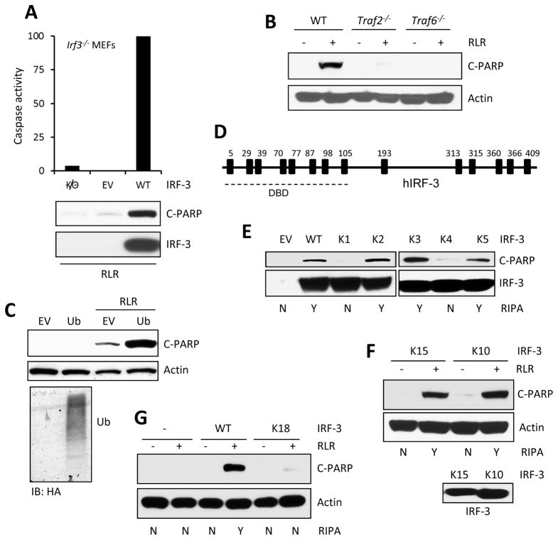
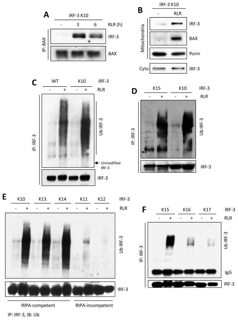
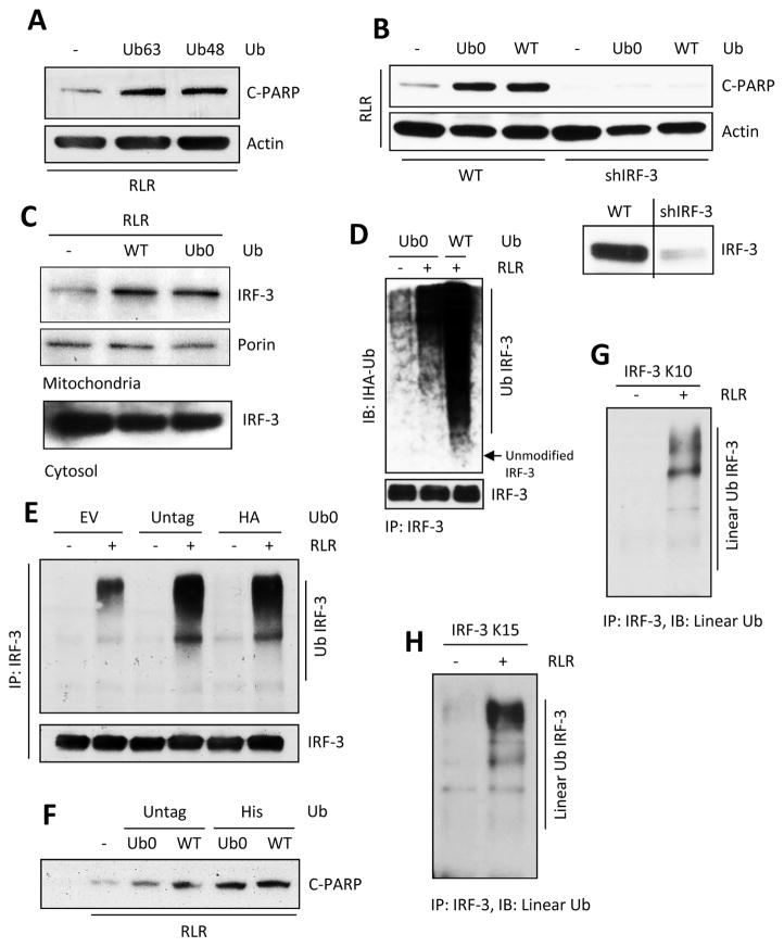
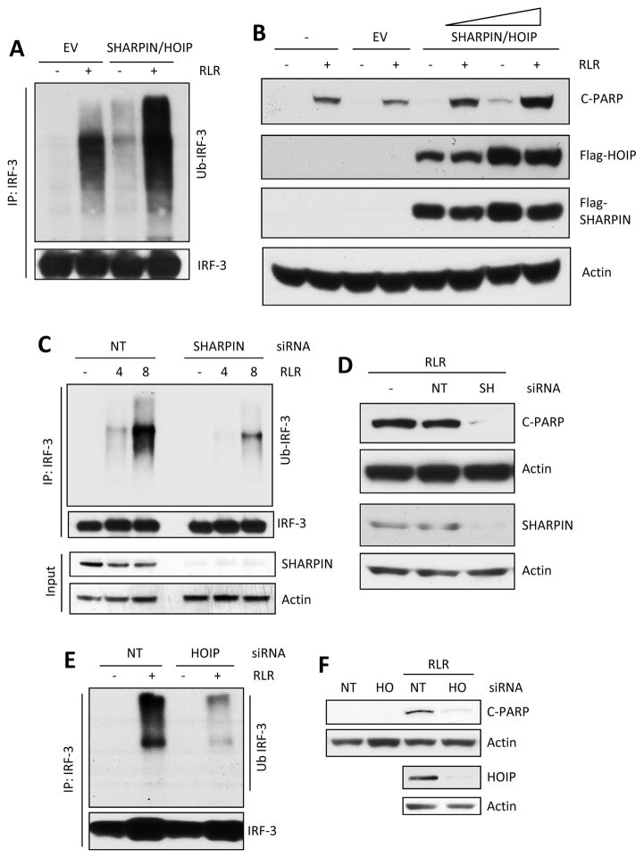
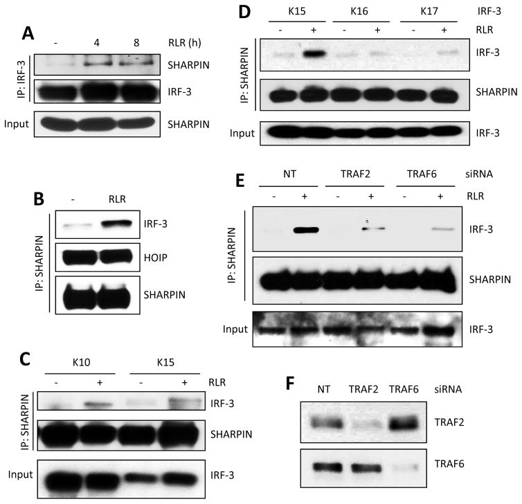
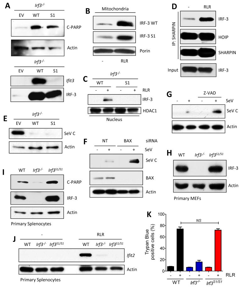
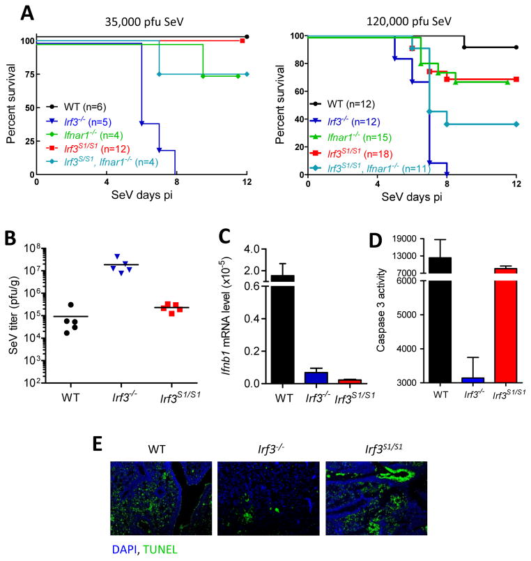
The transcription factor IRF-3 mediates cellular antiviral response by inducing the expression of interferon and other antiviral proteins. In RNA-virus infected cells, IRF-3's transcriptional activation is triggered primarily by RIG-I-like receptors (RLR), which can also activate the RLR-induced IRF-3-mediated pathway of apoptosis (RIPA). Here, we have reported that the pathway of IRF-3 activation in RIPA was independent of and distinct from the known pathway of transcriptional activation of IRF-3. It required linear polyubiquitination of two specific lysine residues of IRF-3 by LUBAC, the linear polyubiquitinating enzyme complex, which bound IRF-3 in signal-dependent fashion. To evaluate the role of RIPA in viral pathogenesis, we engineered a genetically targeted mouse, which expressed a mutant IRF-3 that was RIPA-competent but transcriptionally inert; this single-action IRF-3 could protect mice from lethal viral infection. Our observations indicated that IRF-3-mediated apoptosis of virus-infected cells could be an effective antiviral mechanism, without expression of the interferon-stimulated genes.


Figures

Figure 1
Figure 1. Ubiquitination of IRF-3 promotes RIPA, specific lysines in IRF-3 are required
(A) RIPA was measured by caspase 3 activity or C-PARP concentrations in Irf3−/− MEFs expressing murine IRF-3 (WT) upon poly(I:C) transfection (RLR). (B) C-PARP concentrations in WT, Traf2−/− and Traf6−/− MEFs upon polyI:C transfection (RLR). (C) C-PARP concentrations in HT1080 cells transfected with WT Ub upon poly(I:C) transfection (RLR). The expression of ectopic Ub (HA) in HA-tagged Ub-transfected HT1080 cells (bottom panel). (D) Human IRF-3 and its lysine residues are shown, DBD: DNA binding domain. (E and F) WT or various lysine mutants of human IRF-3 (details in Table S1) were lentivirally expressed in HT1080.shIRF-3 cells; C-PARP was analyzed upon RLR stimulation (polyI:C transfection). (G) C-PARP w concentrations were measured in Irf3−/− MEFs expressing WT or K18 mutant of murine IRF-3 upon poly(I:C) transfection (RLR). EV, empty vector. The results presented here are representatives of at least three biological repeats. Please see Figure S1 for additional data related to Figure 1.
Figure 2
Figure 2. Minimal lysine mutant of IRF-3 is functional in various steps of RIPA and is ubiquitinated on specific lysines
(A) IRF-3 and BAX interaction was analyzed by co-IP assay in IRF-3 K10 mutant expressing cells, upon polyI:C transfection (RLR). (B) Translocation of IRF-3 and BAX to mitochondrial fractions in K10-expressing cells, upon polyI:C transfection (RLR). (C–F) Total ubiquitination of IRF-3 was analyzed for WT or various lysine mutants of IRF-3 in SeV-infected (RLR) cells. The results presented here are representatives of at least three biological repeats. Please see Figure S2 for additional data related to Figure 2.
Figure 3
Figure 3. Unbranched, linear ubiquitination of IRF-3 triggers RIPA
(A) C-PARP was analyzed in HT1080 cells transfected with K63 (Ub63) or K48 (Ub48) mutants of Ub upon polyI:C transfection (RLR). (B) HT1080 (Wt) or HT1080.shIRF-3 cells were transfected with WT or K0 (Ub0) mutant of Ub and C-PARP was analyzed upon polyI:C transfection (RLR); lower panel: IRF-3 expression in HT1080 (Wt) and shIRF-3 cells. (C) Mitochondrial translocation of IRF-3 upon polyI:C transfection (RLR) in WT or K0 (Ub0) Ub transfected cells. (D) Total ubiquitination of IRF-3 in K0 (Ub0) or Wt Ub expressing cells upon polyI:C transfection (RLR). (E and F) Total ubiquitination of IRF-3 (E) and the amounts of C-PARP (F) were analyzed in various Ub transfected cells upon RLR stimulation (E, SeV infection, F, polyI:C transfection). (G and H) Linear ubiquitination of IRF-3 was analyzed for the lysine mutants (as indicated) of IRF-3 upon SeV infection (RLR), using linear ubiquitin specific antibody (Genentech). The results presented here are representatives of at least three biological repeats. Please see Figure S3 for additional data related to Figure 3.
Figure 4
Figure 4. LUBAC is involved in ubiquitination of IRF-3 and RIPA
(A and B) IRF-3 ubiquitination (A, RLR, SeV infection) and RIPA (B, RLR, polyI:C transfection) were analyzed in IRF-3 K10 mutant expressing cells co-transfected with SHARPIN and HOIP plasmids (EV, empty vector). (C and D) IRF-3 ubiquitination (C, RLR, SeV infection) and RIPA (D, RLR, polyI:C transfection) were analyzed in IRF-3 K10 mutant expressing cells transfected with non-targeting (NT) or SHARPIN (SH) siRNA (Thermo Scientific). Silencing of SHARPIN was analyzed by Immuno blot. (E and F) IRF-3 ubiquitination (E, RLR, SeV infection) and RIPA (F, RLR, polyI:C transfection) were analyzed in IRF-3 K10 mutant expressing cells transfected with non-targeting (NT) or HOIP (HO) siRNA (Thermo Scientific). Silencing of HOIP was analyzed by Immuno blot. The results presented here are representatives of at least three biological repeats. Please see Figure S4 for additional data related to Figure 4.
Figure 5
Figure 5. IRF-3 and LUBAC form a complex and this is dependent on TRAFs and specific lysine residues in IRF-3
(A) Co-IP of IRF-3 and SHARPIN in RLR (SeV infection)-stimulated cells at the indicated time. (B–D) SHARPIN and HOIP were co-transfected in IRF-3 mutant (as indicated) expressing cells, and co-IP of IRF-3 and SHARPIN was analyzed upon RLR stimulation (SeV infection). (E) IRF-3 K10-expressing cells were transfected with a non-targeting (NT), TRAF2 or TRAF6 specific siRNA (Thermo Scientific) and co-IP of IRF-3 and SHARPIN was analyzed upon RLR stimulation (SeV). (F) Silencing efficiency of the TRAF2 and TRAF6 specific siRNAs. The results presented here are representatives of at least three biological repeats.
Figure 6
Figure 6. RIPA and transcriptional activities of IRF-3 S1 mutant in vitro and ex vivo
(A) RIPA (C-PARP, top panel) and transcriptional (Ifit3, bottom panel) activities of IRF-3 in RLR (polyI:C transfected) stimulated Irf3−/− MEFs expressing WT or S1 mutant (SS388/90AA) of murine IRF-3. (B and C) RLR-induced translocation of IRF-3 (WT or S1 mutant, expressed in Irf3−/− MEFs) to mitochondria or nucleus; porin and HDAC1 are markers of mitochondrial and nuclear extracts. (D) SHARPIN and HOIP were co-transfected in S1-expressing Irf3−/− MEFs, and co-IP of IRF-3 and SHARPIN was analyzed upon RLR stimulation (SeV infection). (E) Expression of SeV C protein in Irf3−/− MEFs expressing WT or S1 mutant of IRF-3 upon SeV (52 strain) infection. (F) SeV C protein expression in Irf3−/− infected MEFs expressing S1 mutant after transfection of non-targeting or BAX-specific siRNA. (G) SeV C protein expression in Irf3−/− MEFs, expressing S1 mutant, after treatment with caspase inhibitor (Z-VAD). (H) IRF-3 expression in primary MEFs isolated from WT, Irf3−/− and S1 mice analyzed by Immunoblot. (I) Primary splenocytes from mice (genotypes indicated) were infected with SeV (RLR) and analyzed for Ifit2 induction by Immuno blot. (J) Primary splenocytes isolated from the mice (genotypes indicated) were analyzed for RIPA (C-PARP) upon RLR stimulation (polyI:C transfection). (K) Trypan blue-positive dead cells in RLR-stimulated (polyI:C transfected) WT, Irf3−/− and S1 MEFs. The results presented here are representatives of at least three biological repeats. Please see Figure S5 for additional data related to Figure 6.
Figure 7
Figure 7. Genetically targeted Irf3 S1 mice exhibit reduced viral load in lungs and are protected from viral pathogenesis
(A) Mice (genotypes indicated) were intranasally infected with SeV (two different pfu per mouse, as indicated) and monitored for morbidity for the indicated time. B to E is from mice infected with the higher dose of virus. (B) Lungs isolated from SeV-infected mice (genotypes indicated) were analyzed for infectious viral titer by plaque assay. (C) Lung mRNAs from SeV-infected mice were analyzed for IFN-β induction by qRT-PCR. (D) Lung homogenates from SeV-infected mice (genotypes indicated) were analyzed for caspase-3 activity. (E) TUNEL staining from SeV-infected lung sections of the indicated mice. The results presented here are representatives of at least three biological repeats. Please see Figure S6 for additional data related to Figure 7.

References

    1. Belgnaoui SM, Paz S, Samuel S, Goulet ML, Sun Q, Kikkert M, Iwai K, Dikic I, Hiscott J, Lin R. Linear ubiquitination of NEMO negatively regulates the interferon antiviral response through disruption of the MAVS-TRAF3 complex. Cell host & microbe. 2012;12:211–222. - PubMed
    1. Chattopadhyay S, Fensterl V, Zhang Y, Veleeparambil M, Wetzel JL, Sen GC. Inhibition of viral pathogenesis and promotion of the septic shock response to bacterial infection by IRF-3 are regulated by the acetylation and phosphorylation of its coactivators. MBio. 2013a;4:e00636. - PMC - PubMed
    1. Chattopadhyay S, Fensterl V, Zhang Y, Veleeparambil M, Yamashita M, Sen GC. Role of interferon regulatory factor 3-mediated apoptosis in the establishment and maintenance of persistent infection by Sendai virus. Journal of virology. 2013b;87:16–24. - PMC - PubMed
    1. Chattopadhyay S, Marques JT, Yamashita M, Peters KL, Smith K, Desai A, Williams BR, Sen GC. Viral apoptosis is induced by IRF-3-mediated activation of Bax. EMBO J. 2010;29:1762–1773. - PMC - PubMed
    1. Chattopadhyay S, Sen GC. IRF-3 and Bax: a deadly affair. Cell Cycle. 2010;9:2479–2480. - PMC - PubMed